Detection of Specific Microbial Species
Introduction
Detection of specific microbes includes Escherichia coli, Staphylococcus aureus, Pseudomonas aeruginosa, Salmonella, Shigella, Clostridia and Candida albicans as per Indian Pharmacopoeia 2014.
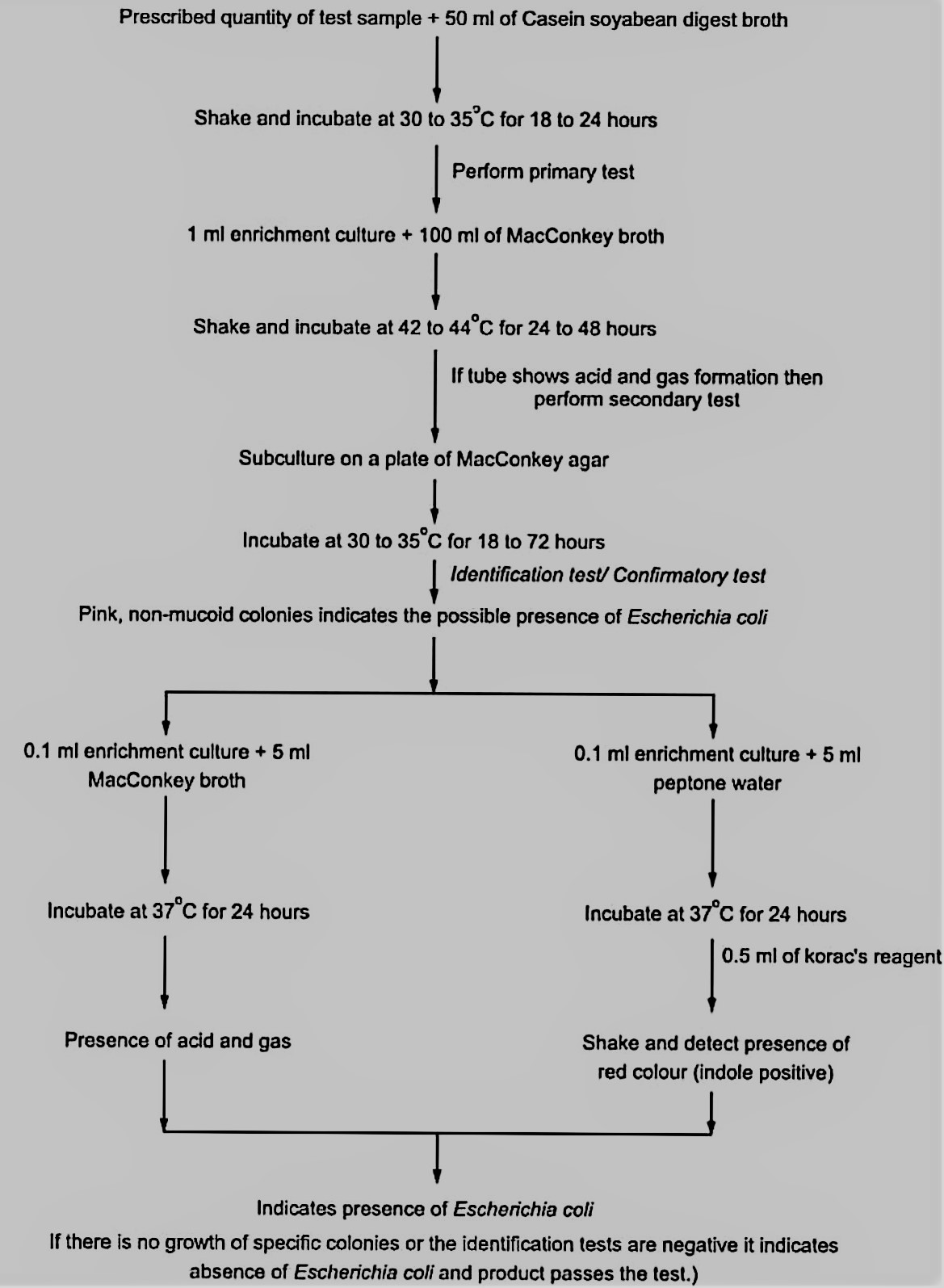
Test for detection of Escherichia coli:
Commonly observed in GIT of mammals and is detectable in feces.
Some strains are harmless and are a part of normal microbial flora of GIT while some strains are pathogenic and produce enterotoxins which cause diarrhoea.
Hence it is necessary to avoid them in pharmaceutical preparations to prevent a chance of infection.

It is a Gram -ve organism.
Non Capsulated bacillus.
Size: 1-3 µm X 0.4 to 0.7 µm.
Facultative anaerobe.
Red or Pink colored colonies on MacConkey agar.
Citrate negative.
Fermentation positive with gas production.
IMViC Test:
Indole: Positive.
Methyl Red: Positive.
Voges-Proskauer: Negative.
Citrate Utilization: Negative.
Salmonella:
More pathogenic than Escherichia coli.
Must be excluded from the pharmaceutical preparations due to potential to cause infection is high.
It is found in the intestines of man, animals and birds, most prominent member is Salmonella typhi.
Gram -ve organism.
Non sporing.
Non acid fast.
Non capsulated bacilli.
Size: 1-3 µm X 0.5 to 0.8 µm.
Motile, Peritrichous except S. gallinarum, S. pullorum.
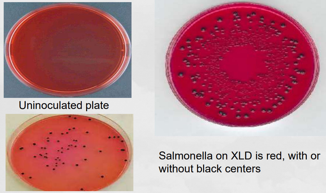
No lactose fermentation: Colonies on MAC and DCA agar are colorless.
Wilson Blair Bismuth Sulphite medium: jet blck colonies with metallic sheen (Production of H2s). (Salmonella paratyphi A produces green colonies as don't produce H2S).
Detection Procedure for Salmonella typhi

Detection of Staphylococcus aureus:
Inhabit skin and nose without any infection but some strains are extreme pathogenic.
Limit tests for this microbe are more likely for topical products.


Gram +ve.
Spherical cocci.
Non motile.
Non sporing.
Grape like clusters.
Some are aerobes and some are facultative anaerobes.
Causes fermentation with no gas production.
Catalase positive.
MR positive.
VP positive.
Indole negative.
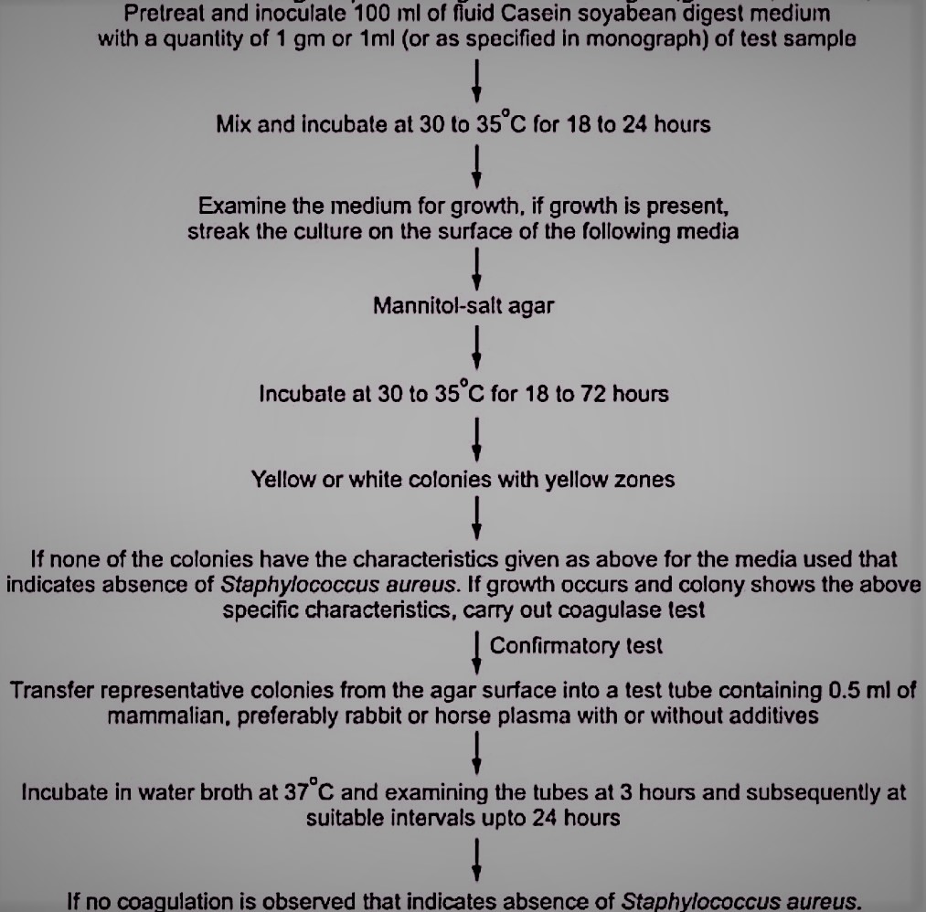
Colonies: Black shiny on Blood Agar, Yellow on MSA agar.
Detection Procedure for Staphylococcus aureus
Detection of Pseudomonas aeruginosa:
Pseudomonas aeruginosa is a potential pathogen causing infections at various sites in the body especially in the eye.
Its strains are found to be resistant to many preservatives at their therapeutic doses hence its a potential problem.
Products for topical application like eye drops are specially tested for presence of Pseudomonas aeruginosa.
Pseudomonas aeruginosa produces a number of water-soluble pigments, including the yellow-green or yellow-brown fluorescent pigment pyoverdin (fluorescein).
When pyoverdin combines with the blue water-soluble pigment pyocyanin, the bright green color characteristic of Pseudomonas aeruginosa is created.
Motile (by single or multiple polar flagella)
gram-negative rods
Obligate (strict) aerobes (most strains)
Oxidase (usually) and catalase positive
Nonfermentative
Chemoheterotrophic
Glucose used oxidatively
On Nutrient agar→ Colonies are surrounded by bluish-green coloration
hemolytic colonies are observed on blood agar
On MacConkey agar- pale yellow colonies i.e. non-lactose fermenters
On selective media “Cetrimide” → pigments are more obvious
Pseudomonas aeruginosa is able to grow at temperatures as high 42 degrees.
MR: Negative (-ve)
VP: Negative (-ve)
Indole: Negative (-ve)
Citrate : Positive (+ve)
Detection Procedure for Pseudomonas aeruginosa

Detection of Clostridia:
Many species of Clostridium are highly toxic.
The toxins produced by Clostridium tetanae are neurotoxins, those produced by Clostridium welchii are histotoxic while those produced by Clostridium difficile are enterotoxins.
The pathogenicity of the organism is due to release of destructive toxins and enzymes.
Very short generation time.
Some prominent organisms which belong to Clostridia are as follows,
Clostridium tetani (Tetanus), Clostridium botulinum (Botulism), Clostridium perfringens (Food Poisoning), Clostridium septicum (Gas Gangrene).
Gram +ve.
Anaerobic.
Capsulated.
Spindle shaped and highly pleomorphic (various shapes and sizes).
Motile: Peritrichate.
Sporulating.
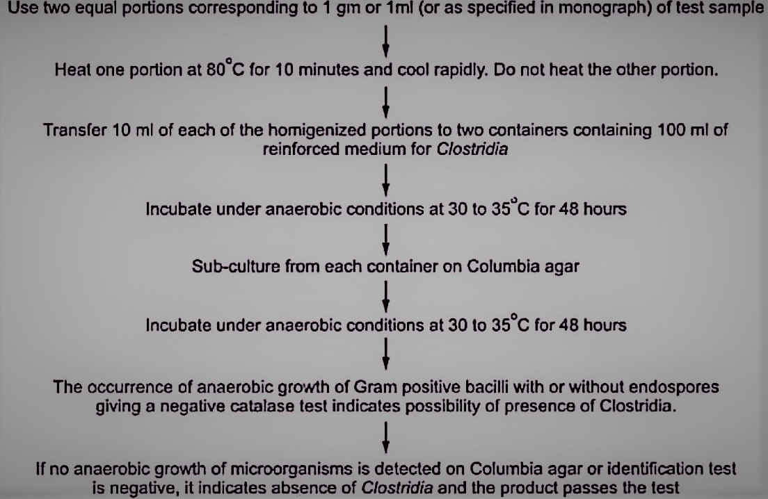
Detection Procedure for Clostridia
Detection of Candida albicans:
It is an optimistic fungus which causes infection of the skin, mucous membrane and internal organs.
It causes candidiasis of Tongue (thrush), Paronychia (Subcutaneous tissues at base of finger nails), Vulvovaginitis (Vagina), Endocarditis (Inflammation of endocardium), Meningitis (Inflammation of brain), Septicaemia (Blood), Bronchocandidiasis (Infection of lungs).
Detection of Candida albicans

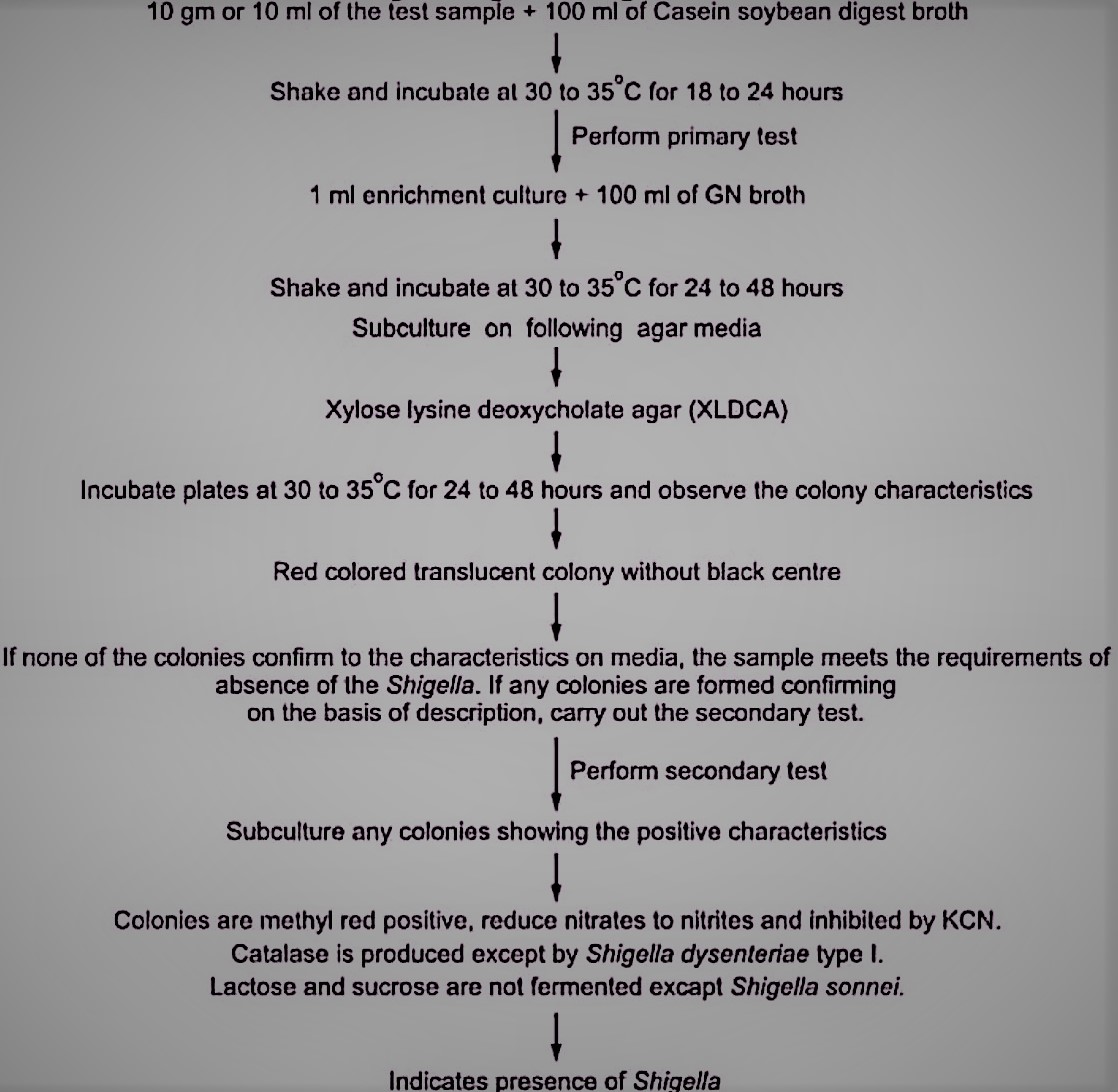
Detection of Shigella:
Shigella is one of the prominent causes of bacillary dysentery, it is characterized by presence of blood and mucus in the watery stool.
Gram -ve.
Non motile.
Non sporing.
Aerobic and some are facultative anaerobic.
No lactose fermentation (Colonies on MAC agar are colorless).
Agar: XLD, DCA, SS.
MR: Positive.
Catalase: Positive.
non sporing
Detection of Shigella.
Labels: Microbiology


<< Home