Connective Tissue.
Tissue level of organization: Connective Tissue.
Introduction:
Groups of cells having similar structure and performing similar functions are called “Tissue.”
Connective tissue is the most abundant tissue in the body.
The connective tissue cells are more widely separated from each other than in epithelial tissues, and the intercellular substance (matrix) is present in considerably larger amounts.
There are usually fibres present in the matrix, which may be of a semisolid jelly-like consistency or dense and rigid, depending upon the position and function of thentissue.
The fibres form a supporting network for the cells to attach to.
Most types of connective tissues have a good blood supply.
Basic functions of Connective Tissue.
Binding
Structural support
Protection
Transport
Insulation.
Important Cells found in Connective Tissue:
Fibroblasts:
Also called “Fibrocytes”.
Large cells.
Produce collagen and elastic fibers.
B) Fat Cells:
Also called Adipocytes.
Occurs as a single cell or in groups.
Size varies as per amount of fat present inside the cell.
C) Macrophages:
These are irregular-shaped cells with granules in the cytoplasm.
Their basic job is eating and digesting foregin substances.
D) Leukocytes:
Normally they are very less in numbers in normal conditions.
Infections trigger their entry in large numbers in the tissue.
E) Mast Cells:
These cells contain large quantities of ‘Histamine” the chemical responsible for many allergies.
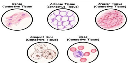
Classification of Connective Tissues:
Loose Connective Tissue:
Areolar Connective Tissue.
Adipose Tissue.
White Adipose Tissue.
Brown Adipose Tissue.
Reticular Connective Tissue.
Dense Connective Tissue:
Fibrous Connective Tissue.
Elastic Connective Tissue.
Cartilage.
Hyaline Cartilage
Fibrous Cartilage
Elastic Fibrocartilage.
Bones:
Compact Bones.
Spongy Bones.
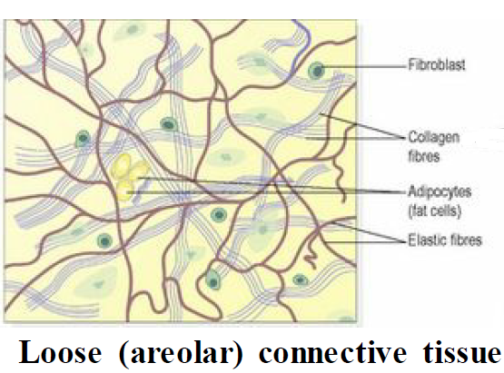
Areolar Connective Tissue:
This is the most common type of connective tissue.
The matrix is semi solid and contains many cells like fibroblasts, macrophages, adipocytes etc.
It is found in almost every part of the body.
It connects and supports other tissues,
It is present under the skin between muscles
Supporting blood vessels and nerves
In the alimentary canal
In glands supporting secretory cells.
Adipose Tissue:
Adipose tissue consists of fat cells (adipocytes), containing large fat globules, in a matrix of areolar tissue.
There are two types: white and brown.

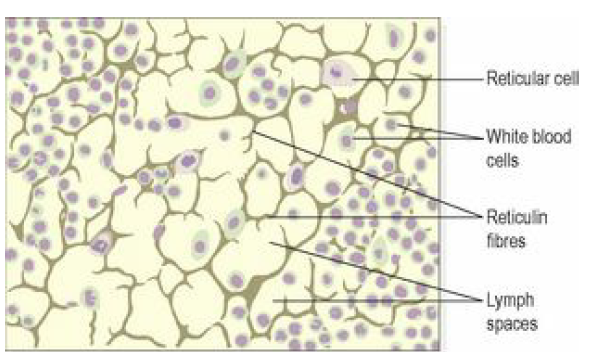
Reticular Connective Tissue:
Also called as “Lymphoid Tissue”.
It contains a semisolid matrix with fine branching reticulin fibres.
It contains reticular cells and white blood cells (monocytes and lymphocytes).
Lymphoid tissue is found in lymph nodes and all organs of the lymphatic system.
Dense Connective Tissue:
Fibrous Connective Tissue:
This tissue is made up mainly of closely packed bundles of collagen fibres with very little matrix.
Fibrocytes (old and inactive fibroblasts) are few in number and are found lying in rows between the bundles of fibres.
Fibrous tissue is found:
forming ligaments, which bind bones together
as an outer protective covering for bone, called periosteum
as an outer protective covering of some organs, e.g. the kidneys, lymph nodes and the brain

Elastic Connective Tissue:
Elastic tissue is capable of considerable extension and recoil.
There are few cells and the matrix consists mainly of masses of elastic fibres secreted by fibroblasts.
It is found in organs where stretching or alteration of shape is required, e.g. in large blood vessel walls, the trachea and bronchi and the lungs.

Cartilage Tissue:
Cartilage is firmer than other connective tissues.
The cells are called chondrocytes and are less in numbers.
Matrix contains a high amount of collagen and elastic fibres.
There are three types:
Hyaline cartilage,
Fibrocartilage
Elastic fibrocartilage.
Hyaline cartilage:
It is a smooth bluish-white tissue.
The chondrocytes are in small groups within cell nests (Pockets).
The matrix is solid and smooth.
Hyaline cartilage provides flexibility, support and smooth surfaces for movement at joints.
It is found:
on the ends of long bones that form joints
forming the costal cartilages, which attach the ribs to the sternum
forming part of the larynx, trachea and bronchi.

Fibrocartilage:
This consists of dense masses of white collagen fibres in a matrix similar to that of hyaline cartilage
The cells widely dispersed.
It is a tough, slightly flexible, supporting tissue found:as pads between the bodies of the vertebrae: the intervertebral discs.

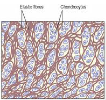
Elastic fibrocartilage:
This flexible tissue consists of yellow elastic fibres lying in a solid matrix.
The chondrocytes lie between the fibres.
It provides support and maintains the shape of, e.g. the pinna or lobe of the ear, the epiglottis and part of the tunica media of blood vessel walls.
D) Bone:
It is the toughest connective tissue.
Bone cells (osteocytes) are surrounded by a matrix of collagen fibres strengthened by inorganic salts, especially calcium and phosphate.
Two types of bone can be identified by the naked eye:
compact bone – solid or dense appearance
spongy or cancellous bone – ‘spongy’ or fine honeycomb appearance.

Commonly Asked Questions.
Define Tissue and explain in detail Connective Tissue.
Write a short note on “Cartilage Tissue”.
Labels: Human Anatomy and Physiology


<< Home