Anatomy & Physiology of Lungs.
Introduction.
A major organ of the respiratory system.
The main function of the lungs is to perform the exchange of oxygen and carbon dioxide.
Two lungs are located in the thoracic cavity above diaphragm.
Gross Anatomy of the Lungs.
The lungs are pyramid-shaped, paired organs that are connected to the trachea by the right and left bronchi.
The diaphragm is the flat, dome-shaped muscle located at the base of the lungs and thoracic cavity.
The lungs are enclosed by the pleurae, which are attached to the mediastinum.
The right lung is shorter and wider than the left lung.
The cardiac notch is an indentation on the surface of the left lung, and it allows space for the heart.
The apex of the lung is the superior region, whereas the base is the opposite region near the diaphragm.
The costal surface of the lung borders the ribs.
The mediastinal surface faces the midline.
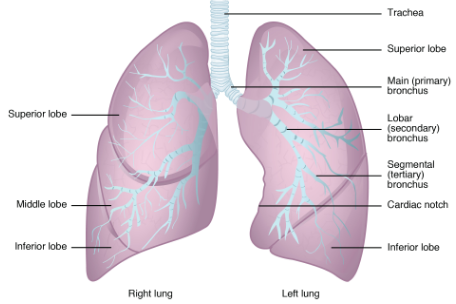
Each lung is composed of smaller units called lobes.
Fissures separate the lobes from each other.
The right lung consists of three lobes: the superior, middle, and inferior lobes.
The left lung consists of two lobes: the superior and inferior lobes.
A bronchopulmonary segment is a division of a lobe, and each lobe houses multiple bronchopulmonary segments.
Each segment receives air from its own tertiary bronchus and is supplied with blood by its own artery.
A pulmonary lobule is a subdivision formed as the bronchi branch into bronchioles.
Each lobule receives its own large bronchiole that has multiple branches.
An interlobular septum is a wall, composed of connective tissue, which separates lobules from one another.
Blood Supply to Lungs.
Lungs receive blood from the pulmonary circulation.
This blood supply contains deoxygenated blood.
The pulmonary artery is an artery that arises from the pulmonary trunk and carries deoxygenated blood to the alveoli.
One arteriole and an accompanying venule supply and drain one pulmonary lobule.
As they near the alveoli, the pulmonary arteries become the pulmonary capillary network.
The pulmonary capillary network consists of tiny vessels with very thin walls that lack smooth muscle fibers.
The capillaries branch and follow the bronchioles and structure of the alveoli.
It is at this point that the capillary wall meets the alveolar wall, creating the respiratory membrane.
Once the blood is oxygenated, it drains from the alveoli by multiple pulmonary veins, which exit the lungs through the hilum.
Nerve Supply to Lungs.
Dilation and constriction of the airway are achieved through nervous control by the parasympathetic and sympathetic nervous systems.
The parasympathetic system causes bronchoconstriction, whereas the sympathetic nervous system stimulates bronchodilation.
Reflexes such as coughing, and the ability of the lungs to regulate oxygen and carbon dioxide levels, also result from this autonomic nervous system control.
Sensory nerve fibers arise from the vagus nerve.
Pleura of the Lungs:
The pleura (plural = pleurae) is a serous membrane that surrounds the lung.
Each lung is enclosed within a cavity that is surrounded by the pleura.
The pleurae consist of two layers.
Visceral Pleura: Internal Layer.
Parietal Pleura: Outer Layer.
The cavity between the Visceral Pleura and Parietal Pleura is called “Pleural Cavity.
Pleural Cavity contains a fluid called “Pleural Fluid”.
The pleural fluid is secreted by mesothelial cells and its main function is as a lubricant which prevents friction between two layers while breathing.

Commonly asked Question.
With a well labelled diagram explain anatomy and physiology of the lungs.
Labels: Human Anatomy and Physiology


<< Home