Tissue is a group of cells having similar structure and function.
Main Tissues found in Human Body:
Epithelial Tissue:
Connective Tissue.
Muscular Tissue.
Nervous Tissue.
Epithelial Tissue:
Also called as “Epithelium”.
It forms covering of organs.
They are derived from all three embryonic layers.
Location of Epithelial Tissue:
Covering of the organs exposed to the outer environment e.g. skin, GI tract, respiratory tract, urinary and reproductive tract etc.
Epithelium tissue forms much of the glandular tissue.
It forms internal lining of the cavities and hollow organs.
Basic Functions of Epithelium Tissue:
Protects body from physical, biological and chemical wear and tear.
Selective transport: Act as gatekeeper as forms covering of the tissues.
Secretion: Secretes mucus and other chemicals.
Absorption: Intestinal columnar epithelium.
Excretion: Sweat Glands.
Structural Features of Epithelium Tissue:
Epithelium tissue is tightly packed.
Non vascular: No blood supply (Receives nutrients and oxygen by diffusion)
Cells shows intracellular junctions for communication.
Cell has three surfaces;
Apical: Upper Surface
Lateral: Side.
Basal: Lower.
The basal layer secretes certain substances like collagen and glycoproteins which forms basal lamina, basal lamina separates epithelium from underlying connective tissue.
The underlying connective tissue secretes and forms reticular lamina.
The reticular lamina and basal lamina together forms the basement membrane which acts as a site of attachment for the epithelium tissue.
Classification of Epithelial Tissues:
Simple Epithelium:
Simple Squamous Epithelium.
Simple Cuboidal Epithelium
Simple Columnar Epithelium.
Stratified Epithelium:
Stratified Squamous Epithelium.
Stratified Cuboidal Epithelium
Stratified Columnar Epithelium.
Transitional Epithelium
Pseudostratified Epithelium.
Simple Squamous Epithelium:
The cells are flat polygonal in shape and are tightly packed.
There is no direct blood supply and the transfer of materials takes place by diffusion from blood vessels of the underlying connective tissue.
The cells show presence of intracellular junctions for communication.
The simple squamous epithelium that lines blood vessels and lymph vessels is specialized for faster chemical transfer and called as “Endothelium”.
The simple squamous epithelium that lines the serous membranes is called as “Mesothelium”, it secretes a secretion called “Serous Fluid” which serves as lubricating fluid and act as a shock absorber.
The alveoli of the lungs where gases diffuse, segments of kidney tubules, and the lining of capillaries are also made of simple squamous epithelial tissue.
Simple Cuboidal Epithelium:
The cells are “Cube” like in structure hence called Cuboidal epithelium.
Width is more as compared to height.
This tissue forms internal linings of Thyroid gland, salivary glands, nephrons etc.
Simple Columnar Epithelium:
The cells have “Column” like in structure hence called Columnar epithelium.
Height is more as compared to width.
This tissue forms internal linings of intestines etc.
They are specialized in functions of absorption and excretion.
Columnar epithelium of intestine has “microvilli” which increases surface area of the cells for absorption.
Goblet cells of intestinal lining and the lining of parts of the respiratory tract such as the trachea have secretory function and secretes mucus.
Ciliated Columnar Epithelium:
Ciliated epithelium consists of columnar cells that have cilia on their apical surface.
Ciliated epithelium lines the nasal cavities, larynx, trachea, and large bronchial tubes.
The cilia sweep mucus, with trapped dust and bacteria from the inhaled air, toward the pharynx to be swallowed.
Another location of ciliated epithelium in women is the lining of the fallopian tubes, the cilia here sweep the ovum, toward the uterus.
Stratified epithelium:
Stratified epithelia consist of several layers of cells of various shapes.
Continual cell division in the lower (basal) layers pushes cells above nearer and nearer to the surface, where they are worn off.
Basement membranes are usually absent.
The main function of stratified epithelium is “Protection”
There are two main types:
Keratinized stratified epithelium
Nonkeratinized stratified epithelium
Keratinised stratified epithelium:
This is found on dry surfaces subjected to wear and tear, i.e. skin, hair and nails.
The surface layer consists of dead epithelial cells that have lost their nuclei and contain the protein keratin.
This forms a tough, relatively waterproof protective layer that prevents drying of the live cells underneath.
Non-keratinised stratified epithelium:
This protects moist surfaces subjected to wear and tear, and prevents them from drying out.
e.g. the conjunctiva of the eyes, the lining of the mouth, the pharynx, the oesophagus and the vagina.
Connective Tissue
Connective tissue is the most abundant tissue in the body.
The connective tissue cells are more widely separated from each other than in epithelial tissues, and intercellular substance (matrix) is present in considerably larger amounts.
There are usually fibres present in the matrix, which may be of a semisolid jelly-like consistency or dense and rigid, depending upon the position and function of thentissue.
The fibres form a supporting network for the cells to attach to.
Most types of connective tissues have a good blood supply.
Basic functions of connective tissue:
binding
structural support
Protection
Transport
Insulation.
Important Cells found in Connective Tissue:
Fibroblasts:
Also called as “Fibrocytes”.
Large cells.
Produce collagen and elastic fibers.
B) Fat Cells:
Also called as Adipocytes.
Occurs as single cell or in groups.
Size varies as per amount of fat present inside the cell.
C) Macrophages:
These are irregular-shaped cells with granules in the cytoplasm.
Their basic job is eating and digesting foregin substances.
D) Leukocytes:
Normally they are very less in numbers in normal conditions.
Infections trigger their entry in large numbers in the tissue.
E) Mast Cells:
These cells contain large quantities of ‘Histamine” the chemical responsible for many allergies.
Classification of Connective Tissues:
Loose Connective Tissue:
Areolar Connective Tissue.
Adipose Tissue.
White Adipose Tissue.
Brown Adipose Tissue.
Reticular Connective Tissue.
Dense Connective Tissue:
Fibrous Connective Tissue.
Elastic Connective Tissue.
Cartilage.
Hyaline Cartilage
Fibrous Cartilage
Elastic Fibrocartilage.
Bones:
Compact Bones.
Spongy Bones.
Areolar Connective Tissue:
This is the most common type of connective tissue.
The matrix is semi solid and contains many cells like fibroblasts, macrophages, adipocytes etc.
It is found in almost every part of the body.
It connects and supports other tissues,
It is present under the skin between muscles
supporting blood vessels and nerves
in the alimentary canal
in glands supporting secretory cells.
Adipose Tissue:
Adipose tissue consists of fat cells (adipocytes), containing large fat globules, in a matrix of areolar tissue.
There are two types: white and brown.
Reticular Connective Tissue:
Also called as “Lymphoid Tissue”.
It contains semisolid matrix with fine branching reticulin fibres.
It contains reticular cells and white blood cells (monocytes and lymphocytes).
Lymphoid tissue is found in lymph nodes and all organs of the lymphatic system.
Dense Connective Tissue:
Fibrous Connective Tissue:
This tissue is made up mainly of closely packed bundles of collagen fibres with very little matrix.
Fibrocytes (old and inactive fibroblasts) are few in number and are found lying in rows between the bundles of fibres.
Fibrous tissue is found:
forming ligaments, which bind bones together
as an outer protective covering for bone, called periosteum
as an outer protective covering of some organs, e.g. the kidneys, lymph nodes and the brain
Elastic Connective Tissue:
Elastic tissue is capable of considerable extension and recoil.
There are few cells and the matrix consists mainly of masses of elastic fibres secreted by fibroblasts.
It is found in organs where stretching or alteration of shape is required, e.g. in large blood vessel walls, the trachea and bronchi and the lungs.
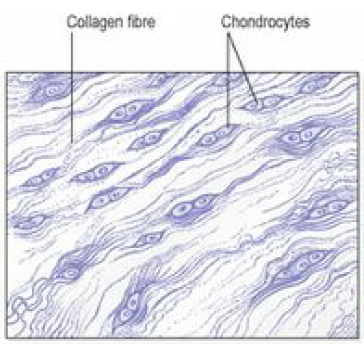
Cartilage Tissue:
Cartilage is firmer than other connective tissues.
The cells are called chondrocytes and are less in numbers.
Matrix contains high amount of collagen and elastic fibres.
There are three types:
hyaline cartilage,
Fibrocartilage
Elastic fibrocartilage.
Hyaline cartilage:
It is a smooth bluish-white tissue.
The chondrocytes are in small groups within cell nests (Pockets).
The matrix is solid and smooth.
Hyaline cartilage provides flexibility, support and smooth surfaces for movement at joints.
It is found:
on the ends of long bones that form joints
forming the costal cartilages, which attach the ribs to the sternum
forming part of the larynx, trachea and bronchi.
Fibrocartilage:
This consists of dense masses of white collagen fibres in a matrix similar to that of hyaline cartilage
The cells widely dispersed.
It is a tough, slightly flexible, supporting tissue found:as pads between the bodies of the vertebrae: the intervertebral discs.
Elastic fibrocartilage:
This flexible tissue consists of yellow elastic fibres lying in a solid matrix.
The chondrocytes lie between the fibres.
It provides support and maintains shape of, e.g. the pinna or lobe of the ear, the epiglottis and part of the tunica media of blood vessel walls.
Bone:
It is the toughest connective tissue.
Bone cells (osteocytes) are surrounded by a matrix of collagen fibres strengthened by inorganic salts, especially calcium and phosphate.
Two types of bone can be identified by the naked eye:
compact bone – solid or dense appearance
spongy or cancellous bone – ‘spongy’ or fine honeycomb appearance.Bone Tissue
Muscle tissue:
This tissue is specialized for contraction and brings movement.
There are three specialized muscular tissues present in the body as follows,
Skeletal Muscles.
Smooth Muscles.
Cardiac Muscles.
Muscle Cells
Skeletal Muscle:
The name is derived as they are attached to the skeleton and are responsible for skeletons movement or say Locomotion.
Also called “Striated muscle” or “Voluntary muscle”.
As they work under our will power they are called “Voluntary muscle”.
On Microscopic examination they appear striated due to precise arrangement of contractile proteins inside the cell.
The plasma membrane and endoplasmic reticulum are special and hence called as “Sarcolemma and Sarcoplasmic reticulum” respectively.
Muscle cells are also called as “Myocytes” , “Muscle Fibers”.
Each muscle fiber contains many rod like structures :Myofibrils.Skeletal Muscle Cell
Skeletal muscle cells are cylindrical in structure and contains many nuclei.
In the cell there is a typical arrangement of proteins responsible for contraction which gives striated appearance.
The impulses for muscle contraction generate at brain or spinal cord and end at neuromuscular junction.
Functions:
Movement.
Body posture.
Regulation of body temperature: Generate heat.
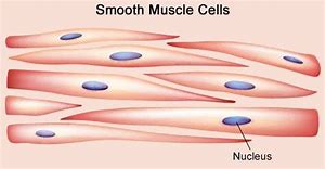
Smooth Muscle:
Smooth Muscle Cells
Also called as “Non Striated Muscles” as don't show striations.
Do not work under will power hence called as “Involuntary Muscles”.
They are present in internal organs hence called as “Visceral Muscles.”
The cells contain single central nucleus and are spindle shaped.
Contractions are slower and more sustained than skeletal muscles.
Functions:
Wall of blood vessels: Regulation of diameter.
Eye: Regulation of pupil size.
Intestine: Peristalsis.
Uterus: Contraction.
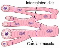
Cardiac Muscle:
Cardiac Muscle Cells
This tissue is only found in myocardium of heart and hence called as “Cardiac Muscle.”
This tissue when observed under microscope shows striations like voluntary muscles but works like involuntary muscle hence it is considered as a special type of muscle.
The cells are branched and has a single nucleus.
The cell membrane at end is folded and fits in matching folds of adjacent cell membrane forming “intercalated discs”.
Intercalated disc is an important feature of cardiac muscle as it makes passage of electric impulses faster.
Heart beats on its own the nerve supply only increases or decreases rate and force of contraction as per need of the situation.
Functions:
Contractions of heart.
Nervous Tissue:
Nervous tissue is a highly specialized tissue in the body and is present in the organs of Nervous system.
Nervous tissue is composed of two types of cells;
Neurons: they initiate, receive, conduct and transmit information.
Neuroglial Cells: They provide support to neurons and also perform some important functions.
Neuroglial Cells:
Neuroglial Cells
Neuroglia word is derived from: Nerve Glue.
Four types of neuroglial cells are found in the central nervous system;
Astrocytes: Forms connection between neurons and blood vessels, imp. Part of BBB (Blood Brain Barrier.)
Microglial Cells: Eat microbes and waste, Protection.
Oligodendrocytes: Insulation and synthesis of Myelin Sheath.
Ependymal cells: Helps in circulation of CSF (CerebroSpinal Fluid).
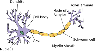
Neuron:
Neuron
They are the main functional cells of the tissue where information is received, analyzed and stored.
Each nerve cell has following important parts;
Dendrites.
Cell Body.
Axon.
Axon terminal.
Dendrites are cell processes that carry impulses towards the cell, a neuron may have many dendrites.
Cell body is also called as “Soma”, contains nucleus and regulates functioning of neurons.
Axon is a long cell process that carries nerve impulses.
Axon terminals are endings of neurons where neurotransmitters are stored to be released in synapse.
Synapse is a minute space between two neurons, the impulse passes through synapse by means of chemicals called “Neurotransmitters”.
Some neurons contains a sheath of a fatty substance around axon called ‘Myelin Sheath” are called as “Myelinated Neurons”.
The neurons which are not covered by Myelin sheath are called as ‘Non Myelinated Neurons”
Myelin sheath is covered by a type of glial cell called “Schwann Cell”.
The groove on Myelinated neuron where myelin sheath is absent and axon is exposed is called as “Node of Ranvier”.
Transfer speed of nerve impulses is faster in myelinated neurons than in unmyelinated neurons.
Questions:
Define and classify tissue.
Give classification of Epithelium Tissue.
Differentiate between Exocrine and Endocrine gland.
Write a short note on,
Muscular Tissue.
Nervous Tissue.
Cartilage Tissue.
Draw well labelled diagram of,
Squamous epithelium.
Cardiac muscle.
Skeletal muscle.
Smooth muscle.
Neuron.
Why nerve impulses travel faster through Myelinated neurons?
In pharmaceutical industries many types of equipments are used for transfer of heat, they can be classified as follows, Heat Exchangers. Heat Interchangers. Heat Exchangers: These devices are used for transferring heat from a fluid (Hot Gas or Steam) to another fluid (Liquid) through a metal wall. Heat Interchangers: These devices are used for transferring heat from a One liquid to another liquid or one gas to another gas through a metal wall. HEAT EXCHANGERS; The equipment used for heat transferring are known as heat exchangers. Some of the processes that involves heat transfer in pharmaceutical industries are: Preparation of starch paste (in steam jacketed kettle). Crystallization. Evaporation. Distillation. Classification of heat exchangers On the basis of transfer of heat, heat exchangers are classified as: Direct transfer type: The hot and cold fluids are separated by a metal wall through which the heat is transferred from hot fluid to cold fluid. E.g. shell and ...
Definition: Biosynthesis of Glycogen from Glucose is called Glycogenesis. Glycogen is synthesized Depending on the demand for glucose and ATP (energy), insulin promotes the glucose conversion into glycogen. Glycogen is the major storage form of carbohydrate in animals similar to starch in plants. It is a homopolymer made up of repeated units of α- D glucose and each molecule is linked to another by 1→4 glycosidic bonds . Once there is a chain consisting of 8 to 10 glycosidic residues in the glycogen fragment, branching begins by 1→6 linkages . Glycogen is stored in liver and skeletal muscles. Location: Cytoplasm of cells in the muscle, liver, and adipose tissue. Steps Involved in Glycogenesis: Glucose is converted into glucose-6-phosphate by the action of glucokinase or hexokinase with conversion of ATP to ADP. Glucose-6-phosphate is converted into glucose-1-phosphate by the action of phosphoglucomutase. Glucose-1-phosphate is converted into UDP-glucose by the actio...
Definition Drying is defined as the removal of liquid from a product usually with application of heat. Rate of Drying Curve. Drying process can be divided into three periods Initial Adjustment Period. Constant drying rate period. First falling drying rate period. Second falling rate period. Initial Adjustment Period (A-B): Also called the “ Heating up” period . In this period the substance gets heat and increases in temperature. Drying has not yet started. Constant drying rate period (B-C): During this period the temperature of the solid and the rate of drying remain constant. The moisture evaporating from the surface is replaced by water diffusing from the interior of the solid at a rate equal t o the rate of evaporation. The moisture content at the end of constant rate (point C) is referred to as the critical moisture content (CMC). At CMC, dry spots start appearing and drying rate starts falling . First falling drying rate period (C-D): This period is also called ...