Digestion is the process of breaking large and complex food molecules into smaller and simple molecules easy for absorption, with help of digestive enzymes and acid.
Like other systems of the body Digestive system also works in coordination with other systems.
The branch of science that deals with the structure, function, diagnosis and treatment of diseases of stomach and intestine is called Gastroenterology.
Digestive System:
It consists of,
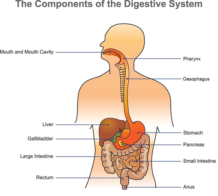
Gastrointestinal Tract (Alimentary Canal):
Mouth,
Pharynx,
Oesophagus,
Stomach,
Small Intestine.
Large Intestine.
Accessory Organs:
Teeth,
Tongue,
Salivary Glands,
Liver,
Gallbladder and Pancreas.
Activities of Digestive System:
Digestive system performs following major activities,
Ingestion: Eating
Movement of Food: Through peristalsis from mouth to anus.
Digestion: Chemical and Mechanical
Absorption:
Defecation: Elimination of undigested waste.
Alimentary Canal:
Alimentary canal is made up of ‘Four Types” of layers throughout its length.
These layers are,
Mucosa.
Submucosa.
Muscularis.
Serosa.
These layers are in continuation with mesentery ( Double fold of peritoneal layer)
Mucosa:
It is also referred to as “Mucous Membrane” as mucous production is one of its main functions.
It is made up of three sub layers,
Epithelium
Lamina propria.
Muscularis mucosa.
Mouth, Phyrenx, Oesophagus and anal canal contains Squamus stratified epithelium.
Stomach and intestines contain Columnar epithelium off which some are modifies to “Goblet Cells” which secrete mucus.
Lamina propria contains some connective tissue and is associated with lymphocytes and forms MALT ( Mucosa associated Lymphoid Tissue) which protects from food borne infections.
Muscularis mucosa is a thin layer of smooth muscles responsible for folding of the stomach and intestinal tissues.
Submucosa:
It is a broad layer of connective tissues consisting of blood vessels and nerves.
It also contains glands that secrete digestive enzymes.
Muscularis:
Third layer is also called “Muscularis externa”.
It is a muscular layer that imparts mechanical digestion.
In the upper and lower part it is made up of skeletal muscles providing voluntary control on digestion and evacuation.
In intestines it is double layer while in stomach it is three layered.
Sclerosa:
Present only on organs inside the abdominal cavity.
Made of a layer of visceral peritoneum and a loose connective tissue.
Main function is to keep organs in position.
Instead of Serosa the mouth, pharynx and oesophagus contains a dense sheet of collagen fibers called adventitia.
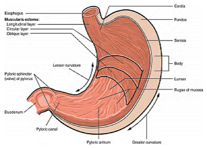
Stomach:
Stomach is present between Oesophagus and First part of small intestine i.e. duodenum.
It is a “J” shaped organ present on the left side of the body and just below the diaphragm.
It is divided into “Four” parts as follows,
Cardia: Superior opening, contains “Cardiac Sphincter”.
Fundus: Superior rounded portion.
Body: Central portion below fundus.
Pylorus: last part, below body, made up of two parts,
Pyloric Antrum:
Pyloric canal: It contains “Pyloric Sphincter”.
Cells of Stomach:
Mucous cells: secrete mucus that protects against stress and acid.
Parietal cells: secrete hydrochloric acid
Chief cells: secrete pepsin, a proteolytic enzyme
G cells: secrete the hormone gastrin
Functions of Stomach:
Reservoir of food.
Secretes gastric acid.
Gastric acid aids in the process of digestion.
Acidic pH in the stomach kills the microbes present in the food.
The churning movements of the stomach cause mixing of food with gastric juices.
The pepsin present in gastric juice helps in protein digestion.
The gastric lipases start digestion of fats.
Acidic drugs like Aspirin get absorbed from the stomach.
Mucosal membrane and mucus of stomach protects linings of stomach from gastric acid.
Cardiac sphincter prevents reflux of acidic contents in oesophagus.
Pyloric sphincter regulates entry of chyme in small intestine and reverse entry into the stomach.
Small Intestine:
It is present between the stomach and large intestine.
It starts at duodenum and ends at ileocaecal junction.
It lies in the abdominal cavity surrounded by large intestine.
Length is 5 Meters and diameter is 1.5 inches.
Provides Chemical as well as mechanical digestion.
Main site for chemical digestion and absorption of food.
Contains “Three” parts,
Duodenum.
First part.
Starts near the pyloric sphincter of stomach.
“C” shaped.
Smallest part is only 25 cm.
Contains openings of bile duct and pancreatic duct.
It has little acidic pH.
Jejunum.
Middle part.
Present between duodenum and ileum.
3 Feet in length.
Ileum.
Last part.
Starts after jejunum and ends at large intestine at “Ileocaecal Junction”.
Length is about 5 Foot and diameter increases from Caecaum to Anus.
In fact length is very less as compared to small intestine but called as Large intestine because of its bigger diameter.
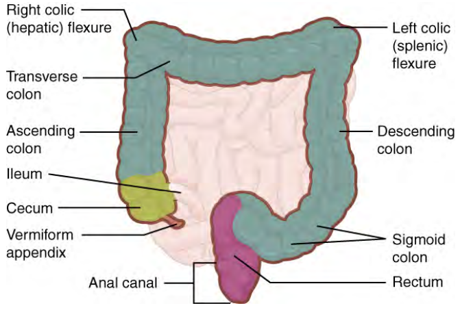
It is present around the small intestine and forms a frame around it.
It is divided into following parts;
Caecum.
Ascending Colon.
Transverse Colon.
Descending COlon.
Sigmoid Colon.
Rectum.
Anal Canal.
Caecum:
First part of large intestine.
Starts at “Ileocaecal Junction”.
Ileocaecal sphincter is present in caecum.
Caecum has a small twisted coiled tubule called “Appendix”.
It ends at “Ascending Colon”.
Ascending Colon:
Remaining part after caecaum is called “Colon”.
Ascending colon is the first part of the colon.
It moves upwards till the lower surface of Liver.
From the liver it turns to the left.
The turn is called “Right Colic Flexure (Hepatic Flexure)”.
Transverse Colon:
It starts from Hepatic Flexure and runs left toward Spleen.
Just below the spleen it turns downwards.
The turn is called “Left Colic Flexure (Splenic Flexure)”.
Descending Colon:
At Splenic Flexure the colon turns and runs downwards till iliac crest.
This part is called a descending colon.
Descending colon ends at Sigmoid Colon.
Sigmoid Colon:
It starts from descending colon and ends in rectum.
Rectum:
Starts from sigmoid colon and ends in anal canal.
13 cm long.
Storage site for fecal material before excretion.
Contains rich blood supply.
Anal Canal:
Last part of GIT that opens in an external environment.
Around 4 cm in length.
Mucous membrane is arranged in folds called “Anal columns” it contains rich blood supply.
Opening to an external environment is called “Anus”.
Anus is guarded by two sphincters,
Internal Anal Sphincter.
External Anal Sphincter.
Internal anal sphincter is made up of smooth muscles hence is involuntary in nature.
External anal sphincter is made up of skeletal muscles hence is voluntary in nature.
These sphincters allow defecation of feces.
Functions of Large Intestine:
Absorption:
Large intestine mainly deals with absorption of water.
It also absorbs many minerals, certain vitamins and alkaline drugs.
Microbial Flora:
Large intestine provides a friendly environment for growth of many microbes and hence is heavily colonized by many bacterias.
These microbes are called “Normal Microbial Flora”.
Normal microbial flora synthesizes certain vitamins like folic acid, cyanocobalamin etc.
The microbial flora also prevents infections from pathogenic microbes found in food.
The microbes when they enter other parts of the body may cause infection e.g. E. coli.
Defecation:
The “mass peristalsis” movement rises in sigmoid colon and pushes fecal material into rectum.
Stretched rectum produces an urge for defecation.
Sphincters are opened and fecal material is expelled out.
Accessory Organs:
An organ that helps with digestion but is not part of the digestive tract.
The accessory digestive organs are the tongue, salivary glands, pancreas, liver, and gallbladder.
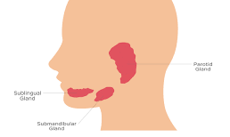
Salivary Glands:
These exocrine glands secrete a secretion called saliva in the oral cavity.
There are 3 pairs of the salivary glands,
Parotid Glands.
Submandibular glands.
Sublingual glands.
Parotid glands:
Largest salivary glands.
Present below the ears.
Secrete saliva in oral cavity through parotid ducts.
Saliva contains large amounts of Salivary amylase.
More fluidity in nature.
Submandibular Glands:
Present below the mandible.
Secrete saliva in oral cavity through submandibular ducts.
Sublingual Glands:
Smallest salivary glands.
Present below the tongue.
Saliva contains less amount of salivary amylase.
Thick in nature.
Saliva:
Secretion of salivary glands is called “Saliva”.
It is colorless.
It is 99.5% water and remaining .5% is inorganic salts, enzymes and waste materials like urea, uric acid etc.
Saliva has a slight acidic pH 6.35 to 6.85.
Saliva contains an enzyme “Salivary amylase (Ptyline)”.
Daily secretion is 1 to 1.5L.
Functions:
Lubrication of the mouth.
Lubrication and moistening of food.
Continuous flushing and cleaning of oral cavity helps checking microbial growth.
Many drugs and waste materials are excreted via saliva.
Salivary amylase causes chemical digestion of starch.
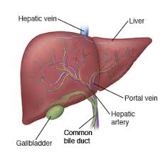
Liver:
Largest gland in the body.
Located on the left side below lungs in the upper abdominal cavity.
Superficially it is covered by peritoneum which separates it into lobes.
Has two main lobes – right and left lobe.
Right lobe is further divided into caudate, and quadrate lobe.
The hepatic blood vessels enter the liver at the porta hepatis.
The gallbladder rests in a recess on the inferior surface of the right lobe.
The structural and functional unit of liver is “Lobule”.
Each liver lobule is hexagonal in shape.
Composed of hepatocyte (liver cell) plates radiating outward from a central vein,
Portal triads are found at each of the six corners of each liver lobule.
Liver sinusoids – enlarged, leaky capillaries located between hepatic plates
Kupffer cells – hepatic macrophages found in liver sinusoids.
Functions of Hepatocytes:
Production of bile
Processing bloodborne nutrients
Storage of fat-soluble vitamins
Detoxification.
Secreted bile flows between hepatocytes toward the bile ducts in the portal triads and is stored in Gallbladder.
Composition of Bile:
A yellow-green, alkaline solution containing bile salts, bile pigments, cholesterol, neutral fats, phospholipids, and electrolytes
Bile salts are cholesterol derivatives that:
Emulsify fat
Facilitate fat and cholesterol absorption
Help solubilize cholesterol
Enterohepatic circulation recycles bile salts
The chief bile pigment is bilirubin, a waste product of heme.
Functions of Liver:
Metabolism:
Carbohydrate Metabolism:
Stores Glycogen.
Gluconeogenesis.
Glycogenolysis.
Fat Metabolism:
Oxidation of fatty acids. (Liver utilizes energy through fat metabolism).
Synthesis of Lipoproteins.
Synthesis of phospholipids and cholesterol.
Converts large amounts of carbohydrates and proteins into fats.
Protein Metabolism:
Parenchymal cells of the liver synthesize plasma proteins.
Deamination of amino acids.
Urea cycle to remove ammonia from the body.
2) Secretion of bile.
3) Storage of many vitamins like fat soluble vitamins and cyanocobalamin.
4) Breakdown of RBCs and defense against microbes by Kuffer cells.
5) Production of heat.
6) Detoxification of drugs and bacterial toxins.
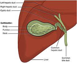
Gallbladder:
It is a thin walled green muscular bag present on the ventral surface of the liver.
8-10 cm long.
Stores and concentrates bile by absorbing its water and ions.
Releases bile via “Cystic Duct” which is connected to “Bile Duct”.
When acidic chyme enters duodenum the “I” cells of small intestine secrete “Cholecystokinin” which stimulate liver for bile production.
Cholecystokinin also causes contraction of the gallbladder releasing bile in duodenum.
Anatomically gallbladder is divided into Fundus, body and neck.
Common diseases of gallbladder include gallstone (Crystallization of cholesterol”, obstructive jaundice.
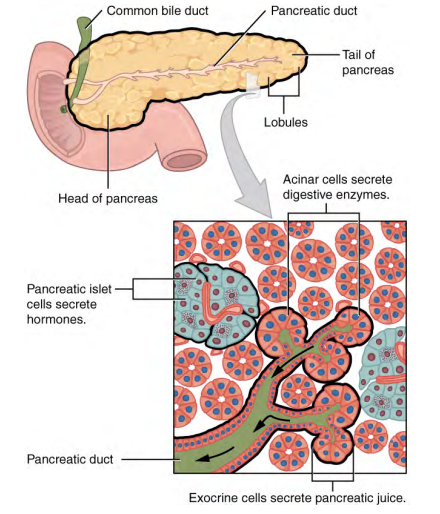
Pancreas:
It is a soft oblong organ present below the stomach.
It is attached to duodenum and ends near spleen.
It has mixed exocrine and endocrine functions.
The pancreas has a head, a body, and a tail.
It delivers pancreatic juice to the duodenum through the pancreatic duct.
The exocrine part of the pancreas arises as little grape-like cell clusters, each called an acinus (plural = acini), located at the ends of pancreatic ducts.
The acini secrete pancreatic juice which contains a large amount of pancreatic enzymes.
The pancreatic duct along with bile duct opens in duodenum in an area called as “Hepatopancreatic ampulla”, this part is guarded by a sphincter called “hepatopancreatic sphincter” which controls entry of bile and pancreatic juices in duodenum.
Islets of Langerhans (EndocrineCells) represents a group of cells that appear surrounded by acini (Exocrine Cells).
Islets of Langerhans contain three types of cells with endocrine function.
Ɑ (Alpha Cells): Secrete “Glucagon”
𝛃 (Beta Cells): Secrete “Insulin”.
𝞭 (Delta Cells): Secrete “Somatostatin”.
Islets of langerhans cells also secrete a local hormone called “Pancreatic Polypeptide”.
Pancreatic Juice:
It's a clear secretion secreted by Pancreas in duodenum.
Pancreas secretes around one liter of pancreatic juice everyday.
It contains mainly water, some salts, sodium bicarbonate and several digestive enzymes.
The proteolytic enzymes are secreted in inactive forms which get activated in the duodenum, while amylases, lipases and others are secreted in active forms.
Pancreatic secretion is controlled by “secretine” a local hormone produced by “S” cells of small intestine.
Hormones of Pancreas:
Endocrine part of pancreas is Islets of Langerhans.
Beta cells of Islets of Langerhans secrete “Insulin” which is responsible for conversion of glucose to glycogen and also for entry of glucose in the cells, hence maintaining the blood sugar levels.
Absence or deficient insulin secretion gives rise to a condition called as “Diabetes mellitus”.
Alpha cells of Islets of Langerhans secrete a hormone called “Glucagon” responsible for conversion of glycogen to glucose, hence providing sugar to the body in fasting conditions.
Delta cells of Islets of Langerhans secretes “Somatostatin” its a local hormone and inhibits secretion of other hormones like “Gastrin, Secretin, Insulin, Glucagon etc.”


<< Home