Special Sense Organs
←
Special Sense Organs:
Introduction:
The special senses include sense of smell, taste, hearing, vision and touch.
A) Sense of smell
-
The sense of smell or olfaction originates in the superior part of the nasal cavity.
-
The nose contains 10-100 millions receptors for the sense of smell.
-
Olfactory receptors react to odorant molecules in inspired air of the nasal cavity.
-
The sense of smell may affect the appetite. If the odours are pleasant the appetite may improve and vice versa.
-
From the olfactory portion of nasal cavity nerve fibres pass through the cribriform plate of the ethmoid bone to the olfactory bulb. From this region nerve fibres are passes to the olfactory area in the temporal lobe of the cerebral cortex. In this area the impulses are interpreted and odour perceived.
-
‘Sniffing’ concentrates more molecules in the roof of the nose. This increases the stimulation of olfactory receptor and the perception of the smell.
B) Sense of taste (gustation)
-
Tongue consists of about 10000 taste buds.
-
The taste buds are present in elevations on the tongue called papillae.
-
There are three types of papillae.
1) Vallate papillae
2) Fungiform papillae
3) Filiform papillae
1) Vallate papillae
-
About 12 large, circular papillae form an inverted V-shaped row at the back side of the tongue.
-
Each of these papillae consist of 100-300 taste buds.
2) Fungiform papillae
-
It is mushroom shaped elevations scattered over the entire surface of the tongue.
-
Each papilla consists of 5 taste buds.
3) Filiform papillae
-
These are thread like structure which increases friction between the tongue and food.
-
It also facilitates movement of food in the oral cavity.

Tastes:
-
Five primary tastes can be distinguished as sour (आंबट) , sweet (गोड), bitter (कडू), salty (खारट) and umami (तुरट) (meaty/savory).
-
All other tastes are a combination of the two or more of the five primary tastes or are associated with olfactory sensation (smell).
-
Certain chemicals stimulate gustation receptors.
-
Nerve impulses are generated and conducted towards the taste area in the parietal lobe of the cerebral cortex where the taste is perceived.
-
The stimulation of different tastes takes place at different parts of the tongue.
1) Sweet and salty taste mainly at the tip of the tongue.
2) Sour taste at the sides of the tongue.
3) Bitter taste at the back of the tongue.
4) Umami taste at the several regions of the tongue.
C) Hearing
-
The organ of hearing is ear.
-
It is supplied by VIIIth cranial nerve (Vestibulocochlear Nerve), which is stimulated by vibrations of sound waves.
-
The ear is divided into three main regions.
1) External (outer ear)
2) Middle ear
3) Internal ear
1) External (Outer ear)
-
The external ear consists of the auricle (pinna), external auditory canal and eardrum (tympanic membrane).
-
The auricle is made up of elastic cartilage which is covered by skin.
-
The external auditory canal is a curved tube about 2.5 cm long and extends from the auricle to the eardrum.
-
The eardrum is a thin, semi transparent part present in between the external auditory canal and middle ear.
2) Middle ear
-
The middle ear is the small cavity present in the temporal bone.
-
The anterior wall of the middle ear contains an opening that directly connects with the nasopharynx called as auditory tube (Eustachian tube).
-
Middle ear consists of three smallest bones present in the body called auditory ossicles.
-
The three bones are the malleus, incus and stapes.
-
The one end of the malleus is attached to the internal part of eardrum.
-
The other end of the malleus articulates with the body of incus.
-
The incus articulates with the head of the stapes.
-
The base of the stapes fits into the oval window.
-
Below the oval window is another opening called the round window.

Internal structure of Ear
3) Internal (Inner) ear
Structurally the internal ear consist of two main divisions,
a) The bony labyrinth :
-
It is a series of cavities present in the temporal bone.
-
It consists of a fluid called perilymph.
-
It is divided into three areas.
i) The semicircular canals -
-
They are anterior, posterior and lateral semicircular canals.
-
Each canal consists of a swollen enlargement at one end called the ampulla.
-
It contains receptors for equilibrium.
ii) The vestibule -
-
It is the oval central portion of the bony labyrinth.
-
It also contains receptors for equilibrium.
iii) The cochlea -
-
It continues with the vestibules.
-
It is a bony spiral canal like a snail shell and makes almost three turns around a central bony core.
-
The cochlea contains receptors for hearing.
b) Membranous labyrinth –
-
These are the series of tubes present inside the bony labyrinth.
-
It is lined by epithelium and contains a fluid called endolymph.
Physiology of hearing:
-
The auricle directs the sound waves into the external auditory canal.
-
These sound waves strike the eardrum and cause the eardrum to vibrate back and forth.
-
The vibration is transmitted from the eardrum to the malleus to the incus and then to the stapes.
-
The vibrations from the middle ear are transferred to the perilymph.
-
From the perilymph the vibrations are transmitted to the endolymph, which leads to the generation of nerve impulses.
-
The generated nerve impulses pass to the auditory portion of the cerebral cortex.
-
These impulses of hearing are interpreted by the brain.
D) Vision
-
The eye is the organ of vision (sight) present in the orbital cavity.
-
The adult eyeball measures about 2.5 cm in diameter.
-
The wall of the eyeball consists of three layers.
1) The superficial fibrous layer
-
It consists of the anterior cornea and posterior sclera.
-
Cornea is transparent layer which covers the coloured iris.
-
The sclera is the ‘white’ of the eye.
-
The sclera covers the entire eyeball except the cornea and gives shape to the eyeball.
-
An opening is present at the junction of the sclera and cornea called scleral venous sinus.
-
A fluid called aqueous humour drains into this sinus.
-
The lacrimal caruncle, is the small, pink, globular nodule at the inner corner of the eye. It is made of skin covering sebaceous and sweat glands.

The front view of Eye
2) The middle vascular layer
-
It consists of three parts: choroid, ciliary body and iris.
-
Choroid consists of various blood vessels which gives nutrients to the posterior surface of the retina.
-
It also contains melanocytes that produce the pigment melanin which prevents reflection and scattering of light within the eyeball.
-
The anterior portion of the choroid becomes the ciliary body.
-
The ciliary body consists of ciliary processes and ciliary muscle.
-
The ciliary processes contain blood capillaries which secretes aqueous humour.
-
The ciliary muscle is a circular band of smooth muscle.
-
Contraction and relaxation of the ciliary muscle changes the shape of the lens for near and far vision.
-
Iris is attached to the ciliary processes, present in between the cornea and the lens.
-
Iris consists of melanin which determines the colour of the eye.
-
The opening in the centre of the iris is called the pupil.
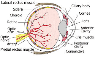
Section of Eyeball
3) The inner nervous layer
-
The inner layer of the eyeball is the retina which covers the posterior part of the eyeball.
-
It is a very delicate structure and responsible for stimulation by light rays.
-
It consists of several layers of retinal neurons.
-
The light sensitive layer consists of sensory receptor cells namely rods and cones.
-
The rods and cones are specialised cells which converts light rays into nerve impulses.
-
Rods stimulate in dim light and do not produce colour vision while cones stimulates in bright light which produce colour vision.
Interior of the eyeball
-
The lens divides the interior of the eyeball into two cavities: the anterior cavity and vitreous chamber.
-
The anterior cavity consists of two compartments namely the anterior and posterior chamber.
-
The anterior chamber present in between the cornea and the iris.
-
The posterior chamber present in between the iris and lens.
-
Both the chambers are filled with a watery fluid that nourishes the lens and cornea called aqueous humour.
-
Vitreous chamber is present in between the lens and the retina.
-
It consists of colourless, transparent, jelly like substance called vitreous body.
-
It maintains intraocular pressure and prevents the wall of the eyeball from collapsing.
E) Touch
-
The skin is the organ of sensation of touch.
-
The skin covers the external surface of the body and it is the largest organ of the body.
-
It consists of two layers: epidermis and dermis and accessory organs like hair, nails, sebaceous glands, and sweat glands.
1) Epidermis
-
The epidermis is made up of keratinized stratified squamous epithelium which produces the protein keratin.
-
Keratin is a tough fibrous protein which protects the skin and underlying tissues from heat, chemicals and microbes.
-
Epidermal cells consist of melanocytes which produce the pigment melanin.
-
Melanin is responsible for skin colour and absorbs ultraviolet (UV) light.
-
In most of the body regions epidermis has four layers: stratum basale, stratum spinosum, stratum granulosum and a thin layer of stratum corneum. This is called ‘thin skin’.
-
In the fingertips, palms and soles, the epidermis has five layers: stratum basale, stratum spinosum, stratum granulosum, stratum lucidum and a thick layer of stratum corneum. This is called ‘thick skin’.
2) Dermis
-
The dermis is made up of connective tissue.
-
Dermal tissue consists of blood vessels, nerves, glands and hair follicles.
-
The dermis is divided into a papillary region and a reticular region.
-
The papillary region consists of areolar connective tissue with fine elastic fibres.
-
The reticular region consists of fine dense irregular connective tissue with collagen fibres and some elastic fibres.
-
It also consists of adipose tissues, hair follicles, nerves, sebaceous (oil) glands and sweat glands.
-
Sebaceous glands of the skin open into the hair follicle.
-
These glands are absent in the palm and sole region.
-
Sebaceous glands secrete some oily secretions called sebum.
-
The sebum keeps skin soft and smooth. It also acts as bactericidal agent which prevents infection by microorganisms.
Functions of the skin
1) The skin regulates body temperature by liberating sweat at its surface and by adjusting the flow of blood in the dermis.
2) The dermis of the skin consists of extensive network of blood vessels and act as blood reservoirs.
3) It provides protection to the body.
4) Cutaneous sensations of the skin are responsible for sensations like touch, pressure, vibration, temperature and pain.
5) Elimination of some substances from the body.
6) The skin acts as absorption medium i.e. the passage of material from the external environment into the body.
7) Synthesis of vitamin D by the ultra violet rays of the sun takes place in the skin.
Physiology of pain
-
Pain serves a protective function by signalling the presence of tissue damaging conditions. Indication of the location of pain may help the underlying cause of disease.
-
Nociceptors (Noci-harmful) are the receptors for the pain which are free nerve endings present in every tissue of the body except the brain.
-
Intense thermal, mechanical and chemical stimuli can activate nociceptors.
-
Tissue irritation and injury release chemicals such as prostaglandins, kinins and potassium ions that stimulate nociceptors.
-
Conditions that generate pain include excess stretching of any structure, prolonged muscular contraction, muscle spasm etc.
QUESTIONS
-
Give the functions of tongue.
-
What are the fundamental sensations of taste?
-
Explain in short V.S. of human skin.
-
Describe the functions of skin.
-
Draw and label diagram of eye.
-
Explain the physiology of pain.
-
Explain mechanism of hearing in short.
-
Explain physiology of taste.
-
Draw and label structure of ear.
-
Write a short note on eye.
Labels: Human Anatomy and Physiology


<< Home