Various Body Fluids and Blood.
Various Body Fluids:
Human body contains two types of body fluids;
Intra cellular fluid
Extracellular fluid
Intra Cellular Fluid:
The fluid of each cell contains its individual mixture of different constituents, but the concentrations of these substances are similar from one cell to another.
The intracellular fluid contains large amounts of potassium and phosphate ions and proteins.
It contains moderate quantities of magnesium and sulphate ions.
The intracellular fluid contains only small quantities of sodium and chloride ions and almost no calcium ions.
Extracellular fluid (ECF):
The extracellular fluid contains large amounts of sodium and chloride ions, reasonably large amounts of bicarbonate ions but only small quantities of potassium, calcium, magnesium, phosphate and organic acid ions are present.
The composition of extracellular fluid is carefully regulated by various mechanisms but especially by the kidneys. This allows the cells to remain continually bathed in a fluid that contains the proper concentration of electrolytes and nutrients for proper cell function.
Examples of ecf includes,
Interstitial fluid (= tissue fluid): Present between the tissues.
Blood Plasma: Present in Blood.
Lymph Plasma: It is present the lymph.
Cerebrospinal Fluid (CSF): It is present inside the brain and spinal cord.
Intraocular Fluid: Present inside the eyeball.
Serous Fluid: Present in serous membranes e.g. Intra pleural fluid, pericardial fluid and peritoneal fluid.
Synovial Fluid: It is present in the synovial joints.
Digestive Fluid: Digestive juices in GIT.
Blood:
Blood is a liquid connective tissue that consists of cells surrounded by an extracellular matrix.
FUNCTIONS OF BLOOD:
Transportation:
Blood transports oxygen from the lungs to the cells of the body and carries carbon dioxide from the body cells to the lungs for exhalation.
It carries nutrients from the gastrointestinal tract to body cells and hormones from endocrine glands to cells throughout the body.
Blood also transports heat and waste products to the lungs, kidneys, and skin for elimination from the body.
Regulation:
Circulating blood helps maintain homeostasis in all body fluids.
Blood plays a role in the regulation of pH through buffers.
It also assists in the adjustment of body temperature.
Protection:
Blood can clot, which protects against its excessive loss after an injury.
In addition, white blood cells protect against disease by carrying on phagocytosis.
Several types of blood proteins, including antibodies, interferons, etc. help protect against diseases.
Physical Characteristics Of Blood:
Blood is thicker than water.
The temperature of blood is about 38C (100.4F), which is slightly higher than normal body temperature.
It has a slightly alkaline pH 7.4.
The color of blood varies.
When saturated with oxygen it is bright red; when unsaturated with oxygen, the blood is dark red to purple.
Blood constitutes about 8 percent of the total body weight.
The blood volume is 5–6 liters in an average-sized adult male and 4–5 liters (1.2 gal) in an average-sized adult female.
Components Of Blood:
Blood is composed of two portions;
Plasma: 55%, A liquid component that contain dissolved substances.
Cell Components: 45%, Blood cells.
Plasma:
It is a straw colored liquid made up of 92% water and 8% dissolved or suspended substances.
The dissolved or suspended substances include;
Plasma Proteins.
Dissolved minerals.
Gases.
Nutrients.
Hormones.
Plasma Proteins:
They constitute around 7% of the plasma.
They are responsible for “Osmotic Pressure” of the blood which keeps the components in plasma in circulation.
All plasma proteins except immunoglobulins are formed in the liver.
Albumins:
They constitute around 60% of total plasma proteins.
Their main function is to create and maintain osmotic pressure.
They also provide binding sites for drugs, hormones and steroids.
Globulins:
Also known as “Antibodies (Immunoglobulins)”.
They are synthesized by Lymphocytes.
They have an important role in immunity that is in protection.
They also provide binding sites to certain hormones and minerals.
Clotting Factors:
The chemicals involved in coagulation of blood are known as clotting factors.
Most common clotting factor is fibrinogen.
The plasma from which clotting factors are removed is called “Serum”.
Electrolytes:
The pH of blood is maintained between 7.35 and 7.45 (slightly alkaline) by an ongoing complicated series of chemical activities, involving buffering systems.
The electrolytes dissolved in plasma has a variety of functions.
Nutrients:
The nutrients absorbed from digestive tracts are present in plasma from where they are transported to various cells.
Nutrients mainly include glucose, amino acids, fatty acids and vitamins.
Waste products:
Urea, creatinine and uric acid are the waste products of protein metabolism.
They are formed in the liver and carried in the blood to the various sites for excretion.
Hormones:
The products of Endocrine glands are poured directly in the blood.
Blood transports hormones to their respective target organs.
Gases:
Oxygen, carbon dioxide and nitrogen are transported around the body dissolved in plasma.
Oxygen is bound to Hemoglobin while most of the Carbon dioxide is available as dissolved bicarbonate ions.
Nitrogen is available in dissolved form but has no physiological role.
Cellular components of blood:
Cellular components of blood includes the blood cells,
Red Blood Cells (Erythrocytes”.
Platelets (Thrombocytes).
White Blood Cells (Leucocytes).
The blood cells are formed in “red bone marrow” present in bones.
In foetus blood cells are formed in the liver and later in bones.
In children the red bone marrow is present in all bones while in adults it is present only in Skull, Ribs, Ending of long bones, Hip Bone, Vertebrae and Sternum.
The formation of blood cells is called “hematopoiesis / hematopoiesis”.
Red Blood Cells:
Also called as “Erythrocytes”.
Bi-concave discs in shape with diameter of 7-8 µm.
They are flexible hence can pass through capillaries by squeezing through.
The plasma membrane is strong and contains specific “Antigens (glycolipids)” which are responsible for blood grouping.
Each RBC contains around 280 million molecules of haemoglobin (Hb).
Mature RBCs lack nucleus and contain maximum amount of Hb.
Mature RBCs also lack mitochondrias and synthesize ATPs required by anaerobic way.
A healthy adult male has about 5.4 million rbcs/ microliter (µL) of blood.
A healthy adult female has about 4.8 million rbcs/ microliter (µL) of blood.
The main function of RBCs is gaseous transportation which is due to the presence of Hb.

Hemoglobin:
Haemoglobin is a large, complex protein containing a globular protein (globin) and a pigmented iron containing a complex called haem.
Each haemoglobin molecule contains four globin chains and four haem units, each with one atom of iron.
Hence a single haemoglobin molecule can carry up to four molecules of oxygen.
An average red blood cell carries about 280 million haemoglobin molecules.

Red Blood Cell Formation:
Also called as Erythropoiesis.
Erythropoietin (EPO) is a hormone produced by the kidney that promotes the formation of red blood cells by the bone marrow.
During foetal stage the blood cell formation takes place in “liver” while before three monthe to birth blood cell formation starts at “Bone marrow”.
The red bone marrow is the major site of blood cell formation.
During childhood almost all the bones contain “red bone marrow”, after attaining adult stage most of red bone marrow is replaced by “yellow bone marrow” which contains adipocytes and lacks the ability to produce the blood cells.
In adults red bone marrow is specifically found in: Skull, Vertebrae, Sternum, Ribs, Pelvic Girdle, Pectoral Girdle and endings of long bones.
Red bone marrow contains “Pluripotent cells” having capacity to get converted into many types of cells.
Pluripotent cells divides and forms “progenitor cells” which are specialized cells that get converted into specific cells.
Progenitor cells divides to develop “Proerythroblasts” which contain nucleus and many ribosomes which are essential for the formation of “hemoglobin”.
Proerythroblast starts producing hemoglobin and then called “Normoblasts”.
Normoblasts are the nucleated RBCs.
Normoblasts when contain approximately 33% hemoglobin nucleus is expelled out and is then called “reticulocyte”.
Reticulocytes get pushed into systemic circulation.
Reticulocytes are premature RBCs which get matured after 1-2 days.
RBC Life Cycle:
Erythropoietin (EPO) is a hormone produced by the kidney that promotes the formation of red blood cells by the bone marrow.
RBCs has a definite lifespan of 120 days.
The old RBCs are destructed in various organs like Spleen, Liver and even in bone marrow.
The spleen is the major organ for RBC destruction.
The fixed macrophages in spleen causes breakdown of RBC the products formed during process are: Globin and Heam.
Globin is a protein fragment.
Heam is a pigment broken down to iron and bile pigments.
Bile pigments are sent to liver and processed and then secreted in bile duct as an element of bile.
Iron is recycled to produce new RBCs.
Blood Group Systems:
RBCs have different types of Antigens present on their surface.
The body synthesises the antibodies against other types of antigens which are not present on the RBCs.
The blood group systems are based on the types of antigen a person is having in RBCs.
Most common blood group systems used are:
ABO System.
Rh System.
ABO System:
The two types of antigens identified present on the RBC surface are Antigen A and Antigen B.
The presence of these antigens gives rise to the blood group system as,
Blood Group A: Having type A Antigen on RBCs.
Blood Group B: Having type B Antigen on RBCs.
Blood Group AB: Having both type A and B Antigens on RBCs.
Blood Group O: No type A & B Antigen on RBCs.
The person of Blood Group A contains antibodies for Antigen B and hence when given blood of Blood Group B shows transfusion reaction.
The person of Blood Group B contains antibodies for Antigen A and hence when given blood of Blood Group A shows transfusion reaction.
The person with Blood Group AB produces no antibodies hence shows no reaction with any blood group and hence called as “Universal Recipient”..
The person with Blood Group O produces both antibodies A & B hence can donate blood to all blood groups and hence is also called as “Universal Donor” however, The person should receive blood from a person having Blood Group O only.
Rh System:
Also called the “Rhesus System.”
It indicates the presence of “Rhesus Factor”. (Rh Factor, D Antigen).
It was first observed in the Rhesus monkey and hence the name was given.
Individuals whose erythrocytes have the Rh antigens (D antigens) are designated Rh +ve while those who lack Rh antigens are designated Rh -ve.
Blood Transfusion:
A transfusion is the transfer of whole blood or blood components into the bloodstream.
It is given to treat low blood volume (Hypovolemia), low Oxygen Carrying capacity of blood (Anaemia), low Platelet count (Thrombocytopenia).
However, in an incompatible blood transfusion, antibodies from recipients blood attacks on donors RBCs causing Hemolysis (breakdown of RBCs).
The free Hb in blood causes kidney problems.
Infected blood products may transmit diseases such as AIDS (Acquired ImmunoDeficiency Syndrome), Hepatitis C & B.
Blood groups compatibility is as given below,

White Blood Cells:
These are the only blood cells which contain “Nucleus” and other cell organelles.
They are termed as “White” as they don't contain “hemoglobin”.
They are the largest blood cells but are only 1% of total blood cells.
NORMAL COUNT: 4500-11000/mm3 of blood.
They are classified as follows,
Classification of WBCs:
Granulocytes:
Neutrophiles.
Eosinophiles.
Basophiles.
Agranulocytes:
Monocytes.
Lymphocytes.
Granulocytes:
The process of formation of granulocytes is called “granulopoiesis”.
All granulocytes contain multilobed nucleus.
Their name represents the dye they take up during staining process,e.g.
Eosinophiles take up “Eosin” the acidic stain and appear “Reddish Orange” in color.
Basophiles take up basic stain “Methylene Blue” and appear bluish.
Neutrophiles take both stains and appear “purple” in color.
Neutrophils:
They are the fastest of all WBCs and reach first at the site of the infection.
They get attracted to the site of infection by chemicals released from damaged tissue parts “Chemotaxis”.
They kill microbes at the site of action by phagocytosis.
Their life span is 6-9 hours.
NORMAL COUNT: 50-70%.

Eosinophils:
These are less active in phagocytosis as compared to the neutrophils.
They are specialized in killing bigger parasites that can not be phagocytosed.
They contain chemicals that can produce inflammation and are often found involved in complications of many allergic conditions like asthma.

Their life span in circulation is 8-10 hrs while in tissues is 8-12 days.
NORMAL COUNT: 1-3%.
Basophils:
They are the largest type of granulocyte.
They can be phagocytic but their main function is in mediation of allergic responses.
They contain chemicals like histamine, heparin , serotonin and are involved in allergic responses viz. Asthma, hay fever and hypersensitivity.
Normally their concentration is very less in circulation but increases significantly in allergic responses.
Their lifespan is 3-10 days.
NORMAL COUNT: 0.5-1%.

Agranulocytes:
They have a large nucleus and no cytoplasmic granules.
They are of two types,
Monocytes.
Lymphocytes
They make up 25-50% of total leucocytes count.
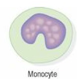
Monocytes:
Largest of WBCs.
Some circulate in blood while some migrate to tissues to become “Macrophages”.
Actively motile and phagocytic.
Macrophages have important functions in inflammation and immunity.
The half-life of blood monocytes is about 1 day, whereas the life span of tissue macrophages is several months or years.
They make up 2-6% of total leucocytes.
Lymphocytes:
Lymphocytes are smaller than monocytes and have large nuclei.
They circulate in the blood and are present in great numbers in lymphatic tissue such as lymph nodes and the spleen.
Although they are formed in bone marrow they get activated in lymphoid tissue.
Lymphocytes are of three types
T-Lymphocytes.
B-Lymphocytes.
Natural Killer Cells
Platelets (Thrombocytes):
These are very small non nucleated cells of blood.
These are formed in the red bone marrow in response to “Thrombopoietin” a substance produced by the kidney in response to low thrombocyte blood count.
They contain many chemicals responsible for blood clotting (Hemostasis).
Lifespan is 8-11 days.
About 1/3 rd of platelets are stored in spleen for emergency condition.
Normal count of platelets is: 150000-450000/ microliter of blood.
Haemostasis:
It is defined as the process of stopping of bleeding and involves many steps like following,
Vasoconstriction:
When damage to the blood vessel takes place many chemicals causing vasoconstriction are released which causes vasoconstriction.
The activated platelets starts aggregating.
Platelet plug formation:
The platelets which are stuck near injury site releases chemicals which attract more platelets.
More platelets comes and adhere with each other forming a plug which prevents further blood loss.
The formed plug is weak hence to make blockade more powerful blood starts clotting.
Coagulation (blood clotting):
It is a complex process involving many steps.
On damage to blood vessel platelets release “Thromboplastin”.
Thromboplastin activates inactive “Prothrombin” to active form “Thrombin” in plasma.
Thrombin activates inactive “Fibrinogen” to active “Fibrin”.
The formed fibrin threads intangle with each other to form a mesh.
The blood cells gets trapped in the fibrin mesh to form a clot.
