CardioVascular System
Introduction:
-
The cardiovascular system consists of the heart, blood vessels and the blood.
-
The hollow muscular pump that circulates blood throughout the body is called as heart.
Anatomy of the heart:
-
The heart is hollow, cone shaped, muscular organ roughly the same size of closed fist of individual.
-
It is about 12 cm long, 9 cm wide and 6cm thick.
-
It weighs about 250 gm in adult female and 300 gm in adult male.
-
It is present in “Mediastinum Region” i.e. on the diaphragm, near the midline of the thoracic cavity, in between the two lungs.
-
About two thirds of the part of the heart is present to the left of the body’s midline.
-
The superior broad part of heart is base and inferior pointed part is apex.
-
The wall of the heart consists of three layers.
-
The Pericardium :( Peri- around)
-
The pericardium membrane surrounds and protects the heart.
-
The pericardium consists of two parts:
-
the fibrous pericardium and
-
the serous pericardium.
The superficial fibrous pericardium is made up of tough inelastic dense irregular connective tissue thus it prevents over distention of the heart.
-
The serous pericardium is the thinner membrane present at the inner side of the fibrous pericardium.
-
It forms a double layer around the heart.
-
The outer is parietal layer and the inner is visceral layer/ epicardium.
-
In between these two layers there is a space called pericardial cavity.
-
This cavity is filled with serous fluid known as pericardial fluid.
-
The Myocardium:
-
It is the middle layer of heart composed of specialized cardiac muscle which is present only in the heart.
-
The myocardium is responsible for pumping action of the heart.
-
The Endocardium:
-
It is the innermost layer of the heart.
-
This lines the chambers and the valves of the heart.
-
The endothelium is continuous with the endothelial lining of the large blood vessels attached to the heart.
Interior of the heart
-
Heart consists of right and left side by the partition in between.
-
Each side is again divided into superior and inferior chambers.
-
Thus the heart consists of four chambers.
-
The two superior chambers are called atria and two inferior chambers are called ventricles.
-
The right atrium and left atrium is separated by a partition called interatrial septum, similarly the right ventricle and left ventricle is separated by the interventricular septum.
-
The valves present between atrium and ventricle are called “Atrioventricular Valves” while the valves present on pulmonary artery and aorta are called “Semilunar Valves”.
-
The blood passes from the right atrium to the right ventricle through a valve called the tricuspid valve.
-
The blood passes from the left atrium to the left ventricle through a valve called bicuspid valve.
-
The blood passes from the right ventricle into the pulmonary artery through the pulmonary semilunar valve.
-
The blood passes from left ventricle into the aorta through the aortic semilunar valve.

Fig.7.1-Interior of Heart
Flow of blood through the heart:
-
The superior and inferior vena cava empties their content into the right atrium.
-
From the right atrium blood passes into the right ventricle through the tricuspid valve.
-
From the right ventricle blood enters into the pulmonary artery which divides into right and left pulmonary arteries. The arteries carry venous blood to the lungs where the blood is oxygenated.
-
Two pulmonary veins from each lung carry oxygenated blood and empty their content into the left atrium.
-
From the left atrium oxygenated blood passes into the left ventricle through the bicuspid valve.
-
From the left ventricle the blood is pumped into the aorta through the aortic valve.
Blood supply to the heart:
-
The heart receives oxygenated blood by the right and left coronary arteries and the venous blood (deoxygenate) is collected into small veins that join to form coronary sinus, which opens into the right atrium.
Cardiac cycle:
-
All the events occurring in the heart with one heartbeat is called cardiac cycle.
-
In a minute about 70 cardiac cycles take place in adults.
-
So the time required for one cardiac cycle is 0.8 sec.
Each cardiac cycle consists of
-
Atrial systole: contraction of the atria called atrial systole which lasts about 0.1 sec.
When there is contraction of atria the blood flows to the ventricles through the atrioventricular valve.
-
Ventricular systole: Contraction of the ventricles is called a ventricular systole which lasts about 0.3 sec.
-
Complete cardiac diastole: It is the relaxation period which lasts about 0.4 sec in which the atria and ventricles are relaxed. As the heart beats faster and faster, the relaxation period becomes shorter and shorter.
Conducting system of the Heart:
-
Small group of specialized neuromuscular cells in the heart initiate and conduct impulses causing synchronized contraction of heart muscle.
The conducting system of heart consist of
-
Sinoatrial node (S.A.node )
-
Atrioventricular Node (A.V.node )
-
Bundle of His
-
Purkinje fibres
-
Sinoatrial node (S.A.node )
-
S.A. node is present at the right atrium just inferior to the opening of superior vena cava S.A. node is also called a pacemaker because it normally initiates the impulses resulting in an action potential. T
-
hese impulses of S.A. node causes atrial contraction
-
Atrioventricular Node (A.V.node ):
-
After the contraction of atria the action potential generated by the SA node, reaches to the AV node located in the septum between the two atria just below the opening of the coronary sinus.
-
There is a gap of 0.1S in initiation of impulse of AV node which results in atrial emptying.
-
Bundle of His:
-
From the AV node the action potential enters in to bundle of his.
-
It is the site where action potential can conduct from atria to ventricle.
-
Purkinje fibres:
-
Finally the purkinje fibres convey the action potential from AV node to apex of heart. Then the ventricle contracts and pumps the blood into the pulmonary artery and the aorta.
Heart sounds:
-
During each cardiac cycle, there are four heart sounds, but in a normal heart, only first and second heart sounds are loud to be heard with the help of a stethoscope.
-
The first sound is “Lubb” which is louder and longer than the second sound. It is caused by the closure of AV valves soon after ventricular systole begins.
-
The second sound is “Dupp” which is short and not as loud as the first. It is caused by the closure of semilunar valve at the beginning of ventricular diastole.
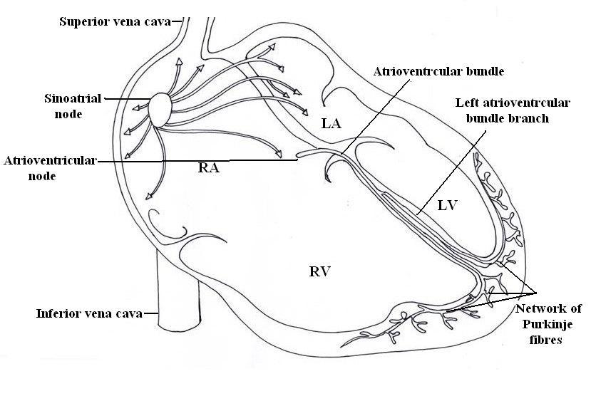
Fig.7.2-Conducting system of Heart
Cardiac output:
-
It is the volume of blood ejected by the ventricle per minute.
-
Cardiac output equals the stroke volume (SV) multiplied by the heart rate (HR).
Stroke volume: It is the volume of blood ejected by the ventricle during each contraction.
CO = SV x HR
CO = 70 ml/ beat x 72 beats/ min.
CO = 5040 ml/ min.
Electrocardiogram:
-
The electrical activity within the body can be detected by attaching the electrodes on the arms and legs (limb leads) and at six positions on the chest (Chest leads).
-
Each limb and chest electrodes records slightly different electrical activity due to difference in its position, by comparing these activities with one another and with normal records, it is possible to determine
-
Abnormality in conducting pathway.
-
Enlargement of heart.
-
If certain region of heart are damaged
-
The cause of chest pain
-
The instrument used is an electrocardiograph and recording of electric signals is an electrocardiogram.
-
The normal ECG shows three waves.
-
The first P wave is a small upward deflection on the ECG.
-
The P wave arises when the impulses from the SA node spreads over the atria.
-
The second wave is the QRS complex starts with a downward deflection continues as a large upright triangular wave and ends as a downward wave.
-
The QRS complex represents rapid spread of impulses from the AV node through the bundle of His and Purkinje fibres.
-
The third wave is dome shaped upward deflection known as the T wave. It indicates the relaxation of ventricular muscles.
Circulation of blood:
-
There are four types of blood circulation in the human body
-
Systemic circulation
-
Pulmonary circulation
-
Coronary circulation
-
Hepatic portal circulation
-
Systemic circulation
-
The left ventricles pumps oxygenated blood into the aorta.
-
From the aorta the blood divides and re-divides into arteries, arterioles and finally the blood capillaries.
-
The exchange of nutrients and gases occurs across the thin capillary walls.
-
The same set of capillaries collects deoxygenated blood from the body cells.
-
They unite to form venules.
-
Different venules comes together to form veins.
-
The veins empty deoxygenated blood either into superior vena cava or inferior vena cava, which opens into the right atrium of the heart.
-
The blood circulation from left ventricle to right atrium is called as systemic circulation.
-
Pulmonary circulation:
-
This is the circulation of blood from the right ventricle of the heart to the lungs and back to the left atrium.
-
Right ventricles pump the blood into the pulmonary artery.
-
It divides into left and right pulmonary artery passes into the left and right lungs.
-
In the lungs these arteries divides and sub divides into smaller arteries, arterioles and capillaries.
-
The exchange of gases takes place between capillaries and the alveoli of the lungs.
-
Two pulmonary veins carry oxygenated blood from each lung and opens in to the left atrium.
-
Coronary circulation:
-
Coronary circulation is the supply of blood to the heart itself. The right and left coronary arteries (Branches of ascending aorta) supply oxygenated blood to the heart.
-
The deoxygenated blood is collected by coronary veins and opens into coronary sinus.
-
Hepatic portal circulation:
-
The venous blood from the digestive organs such as small intestine, stomach, and pancreas is collected by portal vein.
-
It empties its contents into the liver.
-
The portal vein is formed by different veins such as splenic vein from the spleen, inferior mesenteric from rectum and colon, superior mesenteric from small intestine, gastric vein from stomach and cystic vein from gallbladder.
-
In this way, blood with high concentration of nutrients goes in to the liver first.
-
The liver stores some of them and modifies other before they pass into general circulation.
-
The liver also detoxifies harmful substances, such as alcohol, that have been absorbed by the gastrointestinal tract and destroys bacteria by phagocytosis.
-
Hepatic artery supplies oxygenated blood to the liver, thus the oxygenated and deoxygenated blood into the liver is mixed and further collected by hepatic veins which opens into the inferior vena cava.
-
This circulation of blood through the liver is called hepatic portal circulation.
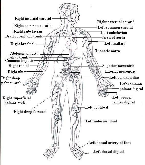
Fig.7.3-Arteries and its branches
Arterial and venous system:
Arterial system:
Aorta-
-
The aorta is the largest artery of the body; the aorta is divided into three divisions.
A) Ascending aorta- It consists of right and left coronary arteries which supplies blood to the heart itself.
B) Arch of aorta
1) Brachiocephalic trunk: It divides into
a) Right common carotid artery: supplies to the right side of head and neck
Which further subdivided in to i) Right internal carotid ii) Right external carotid.
b) Right subclavian artery: Supplies to the right upper limb.
2) Left common carotid artery: supplies to the left side of head and neck
Further subdivided in to i) Left internal carotid ii) Left external carotid
3) Left subclavian artery: Supplies to the left upper limb.
-
There are subclavian arteries.
-
The right subclavian artery arises from the brachiocephalic trunk and left subclavian artery arises directly from arch of aorta.
-
The continuation of subclavian artery in axilla region is called the axillary artery; continuation of the axillary artery in the arm is the brachial artery.
-
At the elbow joint the brachial artery is divided in to a radial and ulnar artery which comes together to form palmar arch; which divides in to palmar digital arteries which supplies blood to the fingers.
C) Descending aorta
1) Thoracic aorta
-
It is about 20 cm long and is a continuation of the arch of aorta.
-
It goes downwards and at the level of the 12th thoracic vertebra it passes through an opening in the diaphragm to form abdominal aorta.
-
The thoracic aorta consists of many paired branches which supplies to the thoracic cavity and the organs within it.

Fig.7.4-Branches of Aorta
2) Abdominal aorta
-
The abdominal aorta is the continuation of thoracic aorta from 12th thoracic vertebra to the 4th lumbar vertebra.
-
The paired branches of abdominal aorta includes the suprarenal, renal and gonadal arteries which supplies to the adrenal glands, kidneys and gonads respectively.
-
At the level of the 4th lumbar vertebra, the abdominal aorta divides into the right and left common iliac arteries.
-
Each common iliac artery is again divided into the internal and external iliac arteries. Internal iliac artery supplies oxygenated blood to the pelvic region.
-
The external iliac artery becomes the femoral artery in the thigh region.
-
The femoral artery passes posterior to the knee to form the popliteal artery which divides into anterior and posterior tibial arteries.
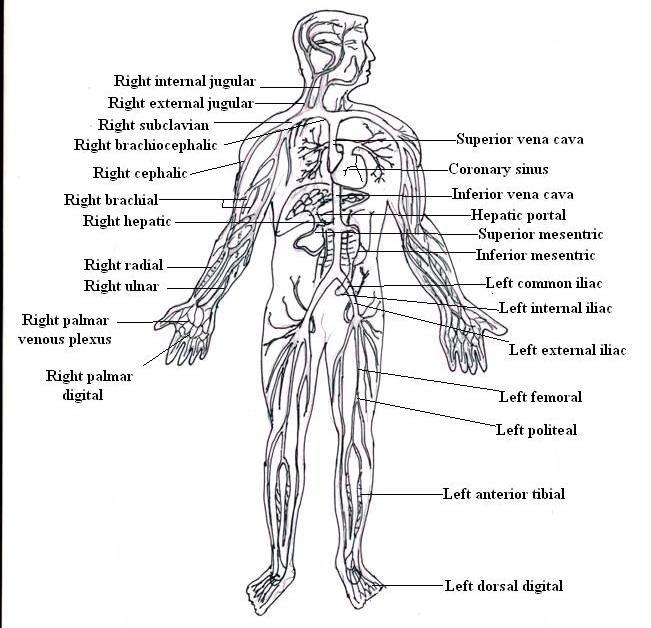
Fig.7.4-Veins and its branches
Venous system
1) Veins of upper limb
-
The radial and ulnar vein unites at elbow to form the brachial vein.
-
It passes superiorly to form axillary vein.
-
At the sternal end of clavicle the axillary vein continues as subclavian vein. The subclavian vein joins with internal jugular vein (which carries blood from the head and neck region) and forms brachiocephalic vein.
-
The brachiocephalic veins of the right and left side comes together to form superior vena cava.
2) Veins of lower limb
-
The anterior and posterior tibial veins at the tibia comes together to form the popliteal vein.
-
It continues as femoral vein in thigh region.
-
The femoral vein passes superiorly to form external iliac vein.
-
It joins with internal iliac vein which carries blood from pelvic organs and forms common iliac vein.
-
The common iliac vein of right and left side comes together to form the inferior vena cava.
3) Veins of head and neck
-
Internal jugular vein and external jugular vein drains the deoxygenated blood from the head and neck.
-
These veins join brachiocephalic vein.
-
The brachiocephalic veins of right and left side comes together to form superior vena cava.
Blood pressure:
-
Pressure exerted by the blood on the walls of blood vessels is called blood pressure.
-
It consists of systolic blood pressure and diastolic blood pressure.
-
Systolic blood pressure is the highest arterial pressure during systole and diastolic blood pressure is the lowest arterial pressure during diastole.
-
The systolic blood pressure in normal adult human is 120 mm of Hg and the diastolic blood pressure is 80 mm of Hg.
-
Pulse pressure is the difference between systolic and diastolic pressure.
-
This pressure provides the information about the condition of the cardiovascular system and is normally about 40 mm of Hg.
Measurement of blood pressure:
-
The device used to measure blood pressure is a sphygmomanometer.
-
It consists of a rubber cuff connected to a rubber bulb and mercury manometer.
-
The cuff is wrapped around an uncovered arm.
-
The stethoscope is placed on the brachial artery.
-
The air is pumped into the cuff with the help of rubber bulb until the brachial artery compress and blood flow stops.
-
Now the air in the cuff is slowly released.
-
It results in the first sound heard through the stethoscope, the reading on the mercury manometer is noted as systolic blood pressure.
-
The air from the cuff is further released and the stage at which sound stops, the reading on the mercury manometer noted as diastolic blood pressure.
-
The normal blood pressure of an adult human is 120/80 mm of Hg (120 is systolic and 80 is diastolic).
Factors modifying blood pressure:
1) Cardiac output
2) Blood volume
3) Peripheral resistance
4) Elasticity of blood vessels
5) Diameter of the lumen of blood vessels
6) Viscosity of blood
1) Cardiac output:
-
It is the volume of blood pumped by the heart per minute, cardiac output depends on the stroke volume when stroke volume increases systolic blood pressure increases. When cardiac output increases both systolic and diastolic pressure increases.
2) Blood volume:
-
Sufficient amount of blood volume is necessary to maintain normal blood pressure. Loss of blood (in haemorrhage) decreases blood pressure.
3) Peripheral resistance:
-
It is the resistance by the walls of blood vessels for the flow of blood due to friction between blood and the walls of blood vessels.
-
The diameters of arteries and veins are large, so their resistance is very small.
-
The smallest vessels like arterioles, capillaries and venules contribute more resistance.
4) Elasticity of blood vessels:
-
Decrease in elasticity result in increase in the blood pressure.
5) Diameter of the lumen of blood vessels:
-
The smaller the lumen of blood vessels, the greater the resistance to blood flow and this increases blood pressure.
-
Larger the lumen of blood vessels, the lesser is the resistance to blood flow and this decreases blood pressure.
6) Viscosity of blood:
-
Higher the viscosity of blood, higher is the resistance which increases blood pressure.
-
The viscosity of blood mainly depends on the concentration of red blood cells in the plasma volume.
Cardiovascular disorders
These are divided into
A) Disorders of the heart.
B) Disorders of the blood vessels.
C) Disorders of the heart
1) Cardiac failure:
-
In this the heart is failing to maintain the circulation of sufficient blood to meet the needs of the body.
-
In cardiac failure left ventricular failure is more common than right, because of the greater workload of the left ventricle.
2) Angina pectoris:
-
Partial obstruction of blood flow in the coronary arteries leads to reduced blood flow to the heart. It causes severe chest pain, which may also passes to the arms, neck and jaw.
-
Angina pectoris is a symptom of imbalance in oxygen demand and supply to myocardium.
3) Myocardial infarction:
-
A complete obstruction of blood flow in the coronary artery results in myocardial infarction commonly known as a heart attack, because of the interrupted blood supply there is the death of the heart tissue.
4) Rheumatic heart disease:
-
It is an autoimmune disease caused due to streptococcal infection.
-
The antibodies produced to fight the original infection damage the connective tissues of the heart.
5) Infective endocarditis:
-
Pathogenic organisms such as bacteria or fungi in the blood may settle at any part of the heart, the most common sites are on or near the heart valves.
6) Cardiac arrhythmias:
-
The rhythm of heartbeats is produced by the SA node.
-
The arrhythmia is an abnormal rhythm as a result of defect in the conduction system of the heart.
-
Symptoms include chest pain, shortness of breath, dizziness.
7) Congenital heart disease:
-
Congenital heart disease is abnormalities in the heart and blood vessels at birth.
-
Most of the defects are in variation in the formation of the septum dividing the heart into right and left side.
B) Disorders of the blood vessels
1) Arteriosclerosis:
-
Thickening of the walls of the arteries and loss of elasticity is called arteriosclerosis.
2) Atherosclerosis:
-
It is nothing but the formation of atherosclerotic plaques in the walls of large and medium sized arteries due to high blood levels of cholesterol.
3) Aneurysm:
-
It is an abnormal local dilation of artery.
4) Embolus:
-
A blood clot, air bubble or fat transported by the bloodstream and block the small vessels.
5) Thrombus:
-
Clotting in the blood vessel which obstructs a blood vessel at the point where it is actually formed.
6) Phlebitis:
-
(Phleb- Vein) It is an inflammation of a vein.
7) Thrombophlebitis:
-
Inflammation of vein due to clot formation.
8) Varicose veins:
-
Varicose vein is a condition in which leaky venous valves can cause veins to become dilated and twisted in appearance.
-
The leaking venous valves allow the backflow of blood which causes pooling of blood.
-
This in turn, creates pressure that distends the vein.
-
As a result the affected veins and the tissue around it may become inflamed and painful.
QUESTIONS
-
Draw a well labelled diagram of L.S. of human heart.
-
What is blood pressure? How it is recorded?
-
Mention any seven cardiovascular disorders.
-
Describe how circulation of blood taken place through heart chambers.
-
Define blood pressure. Explain all factors that modify blood pressure.
-
Describe in brief cardiac cycle.
-
Describe in brief systemic circulation.
-
Define: i) Myocardial infarction ii) Cardiac output iii) Pulse iv) Stroke volume
-
Describe conducting system of heart.
-
Explain the anatomy of the heart.