Introduction:
It differentiates between the physical and chemical properties of two different groups of organisms based on cell-wall characteristics.
To perform this, it requires the use of or more than one stains.
It is further classified into two types as;
Gram Staining.
Acid Fast Staining.
Gram Staining:
This separates bacterias into two major groups,
Gram +ve Bacteria.
Gram -ve Bacteria.
This technique was invented by Danish Dr Hans Christian Gram in 1884.
Principle:
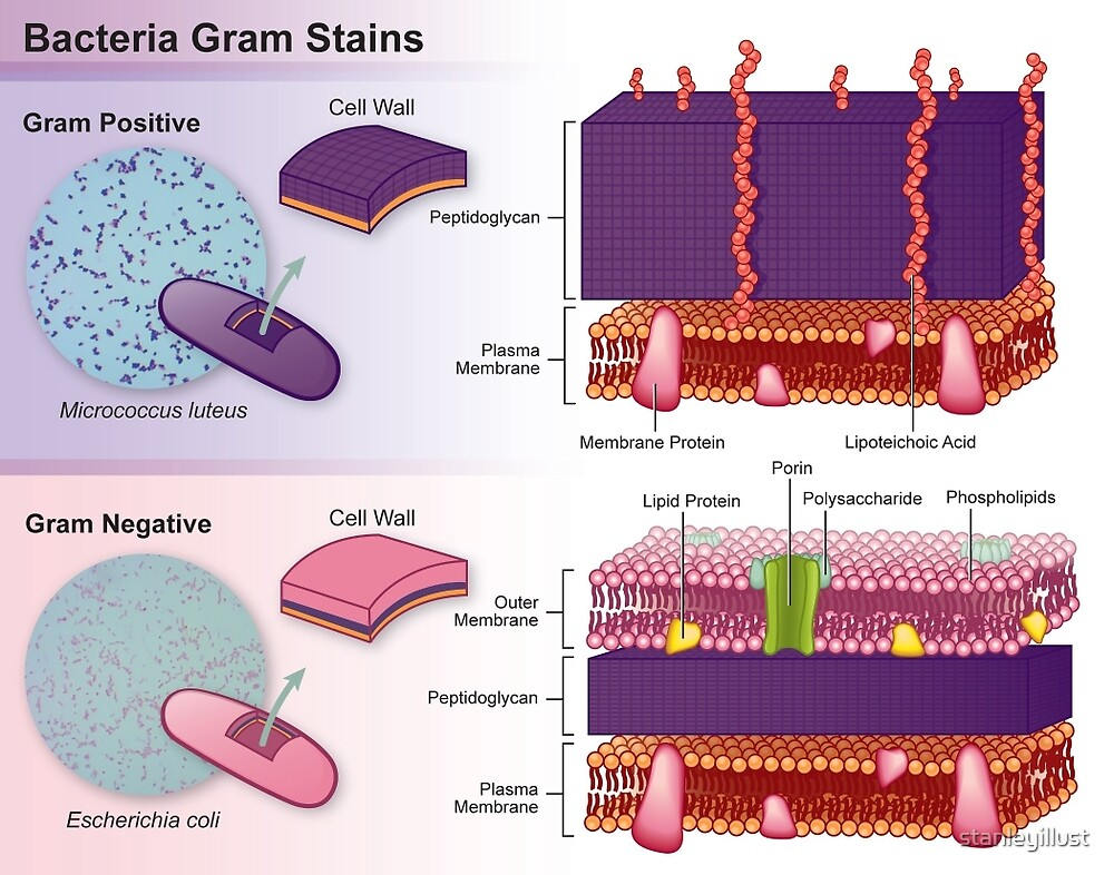
The Gram stain technique is based on the differential structure of the cellular membranes and cell walls of Gram +ve and Gram -ve bacterias.
Gram +ve Bacteria:
Gram-positive organisms contain a highly cross-linked layer of peptidoglycan that retains the primary dye, crystal violet (CV), following the application of the mordant, iodine (I).
The iodine and crystal violet form a complex (CV-I) within the peptidoglycan.
When decolorizer is applied to the cells, the CV-I complex remains within the cell, making it appear dark purple to blue.
Gram -ve Bacteria:
The gram-negative organisms do not contain a thick cross-linked layer of peptidoglycan, which is loosely distributed.
Following the application of the crystal violet and iodine, the CV-I complexes are not trapped within the peptidoglycan.
Application of the acid-alcohol decolorizer dehydrates the outer cellular membrane, leaving holes in the membrane and effectively washing or removing the CV-I complex from the cells.
The cells appear colorless.
To make the colorless cells visible, a secondary stain, safranin, is applied, making the gram-negative cells pink.
Gram Stain Reagents:
Primary stain: Crystal Violet.
Gram’s iodine: (Potassium Iodide +Iodine).
Decolorizer: 50 mL acetone and 50 mL ethanol.
Counterstain: Safranin.
Procedure:
Prepare and fix the specimen to the microscope slide before staining.
Cover the smear with crystal violet, the primary stain, for 20 seconds.
Gently rinse off the stain with water.
Cover the smear with Gram’s iodine for 1 minute.
Pour off the excess Gram’s iodine.
Run the decolorizer over the smear until the solution appears clear.
Gently rinse with water.
Cover the smear with safranin, the secondary or counterstain, for 20 seconds.
Gently rinse the stain with water.
Blot dry with highly absorbent paper and observe under a microscope.

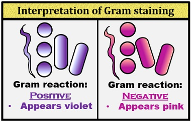
Observation:
Gram-positive: Blue/Purple Color
Gram-Negative: Red/Pink Color
Examples:
Gram-Positive: Streptococcus, Staphylococcus, Corynebacterium, Listeria, Bacillus, Clostridium, etc.
Gram-Negative: E. coli, Salmonella Typhi, Shigella spp, Pseudomonas aeruginosa, Neisseria gonorrhoeae, Chlamydia trachomatis, Yersinia pestis, etc.
Acid Fast Staining:
Acid-fast staining was first introduced by a scientist Paul Ehrlich in the year 1882.
Later, this was modified by Ziehl and Neelson in the year 1883.
Thus, acid-fast staining is sometimes called Ziehl and Neelson staining.
It is a type of differential staining method, which is used to differentiate between the acid-fast and non-acid fast bacteria.
Objective:
To differentiate Mycobacterium species from other species of bacteria.
Differentiate between the acid-fast and non-acid fast bacteria.
Principle:
Mycobacterium does not bind readily to simple stains and therefore the use of heat along with carbol-fuchsin and phenol allows penetration through the bacterial cell wall for visualization.
Mycobacterium cell wall contains high lipid content made up of mycolic acid on its cell wall making it waxy, hydrophobic, and impermeable.
These are ß-hydroxycarboxylic acids made up of 90 carbon atoms that define the acid-fastness of the bacteria.
Use of Carbol-fuchsin which is basic strongly binds to the negative components of the bacteria which include the mycolic acid and the lipid cell wall. The addition of acid alcohol along with the application of heat forms a strong complex that can not be easily washed off with solvents.
The acid-fast bacilli take up the red color of the primary dye, carbol-fuschin.
While non-acid-fast bacteria easily decolorize on the addition of the acid-alcohol and take up the counterstain dye of methylene blue and appear blue
This technique has been used in the identification of Mycobacterium tuberculosis and Mycobacterium leprae.
Reagents:
Primary Stain: Carbol-fuchsin.
Decolorizer: acid-alcohol or 20% Sulphuric acid.
Counterstain: Methylene Blue or Malachite green.
Procedure :
On a clean sterile slide, make the smear of the sample culture and heat fix the smear over blue heat.
Over the smear, pour and flood the smear with carbol fuchsin and heat gently until it produces fumes.
Allow it to stand for 5 minutes and wash it off with gently flowing tap water.
Add decolorizer and leave it for 1-2 minutes. Repeat this step until the smear appears pink in color.
Wash off the acid with water.
Flood the smear with methylene blue dye and leave it for 2-3 minutes and wash with water.
Air dry and examine the stain under the oil immersion lens.
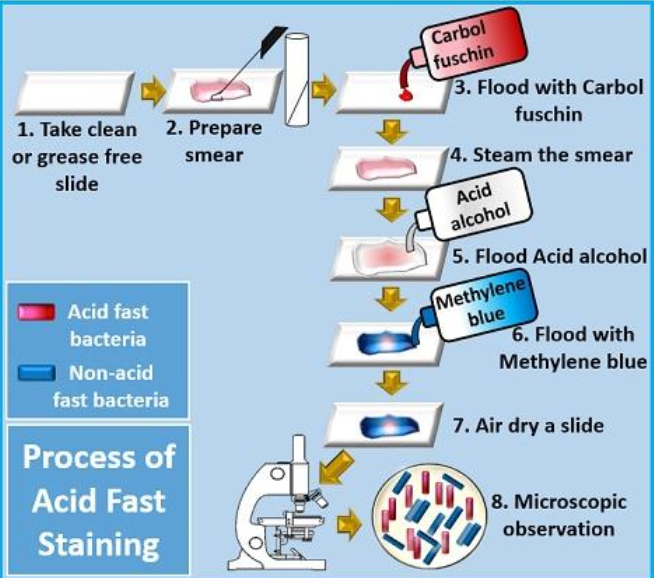
Results:
Acid-fast bacteria retain the primary dye, carbol-fuschin, and stain pink.
Non-acid fast bacteria take up the counterstain methylene blue dye and appear blue.

Applications:
Used for examination and identification of Mycobacterium species.
Used to differentiate between acid-fast and non-acid fast bacilli
Used for the identification of some fungal species such as Cryptosporidium.