Cardiovascular System: Anatomy of Heart & Cardiac Cycle.
Introduction:
The cardiovascular system consists of the heart, blood vessels and the blood.
The hollow muscular pump that circulates blood throughout the body is called the heart.
Anatomy of the heart:
The heart is a hollow, cone shaped, muscular organ roughly the same size as the closed fist of an individual.
It is about 12 cm long, 9 cm wide and 6cm thick.
It weighs about 250 gm in adult females and 300 gm in adult male.
It is present in the “Mediastinum Region” i.e. on the diaphragm, near the midline of the thoracic cavity, in between the two lungs.
About two thirds of the part of the heart is present to the left of the body’s midline.
The superior broad part of the heart is base and the inferior pointed part is apex.
The wall of the heart consists of three layers.
Pericardium.
Myocardium.
Endocardium.
The Pericardium :( Peri- around)
The pericardium membrane surrounds and protects the heart.
The pericardium consists of two parts:
the fibrous pericardium and
the serous pericardium.
The superficial fibrous pericardium is made up of tough inelastic dense connective tissue thus it prevents over distention of the heart.
The serous pericardium is the thinner membrane present at the inner side of the fibrous pericardium.
It forms a double layer around the heart.
The outer is the parietal layer and the inner is the visceral layer/ epicardium.
In between these two layers there is a space called pericardial cavity.
This cavity is filled with serous fluid known as pericardial fluid.
The Myocardium:
It is the middle layer of the heart composed of cardiac muscles.
The myocardium is responsible for pumping the heart.
The Endocardium:
It is the innermost layer of the heart.
This lines the chambers and the valves of the heart.
The endothelium is continuous with the endothelial lining of the large blood vessels attached to the heart.
Interior of the heart
Heart consists of the right and left side chambers by the partition in between.
Each side is again divided into superior and inferior chambers.
Thus the heart consists of four chambers.
The two superior chambers are called atria and two inferior chambers are called ventricles.
The right atrium and left atrium is separated by a partition called interatrial septum, similarly the right ventricle and left ventricle is separated by the interventricular septum.
The valves present between the atrium and ventricle are called “Atrioventricular Valves” while the valves present on the pulmonary artery and aorta are called “Semilunar Valves”.
The blood passes from the right atrium to the right ventricle through a valve called the tricuspid valve.
The blood passes from the left atrium to the left ventricle through a valve called a bicuspid valve.
The blood passes from the right ventricle into the pulmonary artery through the pulmonary semilunar valve.
The blood passes from the left ventricle into the aorta through the aortic semilunar valve.
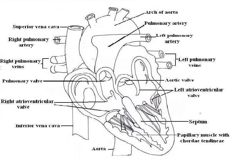
Fig.7.1-Interior of Heart
Flow of blood through the heart:
The superior and inferior vena cava empties their content into the right atrium.
From the right atrium blood passes into the right ventricle through the tricuspid valve.
From the right ventricle blood enters into the pulmonary artery which divides into right and left pulmonary arteries. The arteries carry venous blood to the lungs where the blood is oxygenated.
Two pulmonary veins from each lung carry oxygenated blood and empty their content into the left atrium.
From the left atrium oxygenated blood passes into the left ventricle through the bicuspid valve.
From the left ventricle the blood is pumped into the aorta through the aortic valve.
Blood supply to the heart:
The heart receives oxygenated blood by the right and left coronary arteries and the venous blood (deoxygenate) is collected into small veins that join to form the coronary sinus, which opens into the right atrium.
Cardiac Cycle:
All the events occurring in the heart with one heartbeat are called a cardiac cycle.
In a minute about 70 cardiac cycles take place in adults.
So the time required for one cardiac cycle is 0.8 sec.
Each cardiac cycle consists of
Atrial systole: contraction of the atria called atrial systole which lasts about 0.1 sec.
When there is contraction of atria the blood flows to the ventricles through the atrioventricular valve.
Ventricular systole: Contraction of the ventricles is called a ventricular systole which lasts about 0.3 sec.
Complete cardiac diastole: It is the relaxation period which lasts about 0.4 sec in which the atria and ventricles are relaxed. As the heart beats faster and faster, the relaxation period becomes shorter and shorter.
Commonly Asked Questions:
Draw a well labelled diagram of the interior of the heart.
With a well labelled diagram describing the structure of the human heart.
Write in short about blood flow through the heart.
Write a short note on “Cardiac Cycle”.