Cardiovascular System: Conduction System, ECG and Types of Blood Circulation.
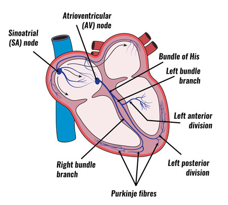
Conducting system of the Heart:
Small group of specialized neuromuscular cells in the heart initiate and conduct impulses causing synchronized contraction of heart muscles.
The conducting system of heart consist of,
Sinoatrial node (S.A.node )
Atrioventricular Node (A.V.node )
Bundle of His
Purkinje fibres
Sinoatrial node (S.A.node ):
S.A. node is present at the right atrium just inferior to the opening of superior vena cava.
S.A. node is also called a pacemaker because it normally initiates the impulses resulting in an action potential.
These impulses of the S.A. node cause atrial contraction .
Atrioventricular Node (A.V.node ):
After the contraction of the atria the action potential generated by the SA node, reaches to the AV node located in the septum between the two atria just below the opening of the coronary sinus.
There is a gap of 0.1S in initiation of impulse of AV node which results in atrial emptying.
Bundle of His:
From the AV node the action potential enters into a bundle of his.
It is the site where action potential can conduct from atria to ventricle.
Purkinje fibres:
Finally the purkinje fibres convey the action potential from the AV node to the apex of the heart.
Then the ventricle contracts and pumps the blood into the pulmonary artery and the aorta.
Heart sounds:
During each cardiac cycle, there are four heart sounds, but in a normal heart, only the first and second heart sounds are loud enough to be heard with the help of a stethoscope.
The first sound is “Lubb” which is louder and longer than the second sound. It is caused by the closure of AV valves soon after ventricular systole begins.
The second sound is “Dupp” which is short and not as loud as the first. It is caused by the closure of the semilunar valve at the beginning of ventricular diastole.
Cardiac output:
It is the volume of blood ejected by the ventricle per minute.
Cardiac output equals the stroke volume (SV) multiplied by the heart rate (HR).
Stroke volume: It is the volume of blood ejected by the ventricle during each contraction.
CO = SV x HR
CO = 70 ml/ beat x 72 beats/ min.
CO = 5040 ml/ min.
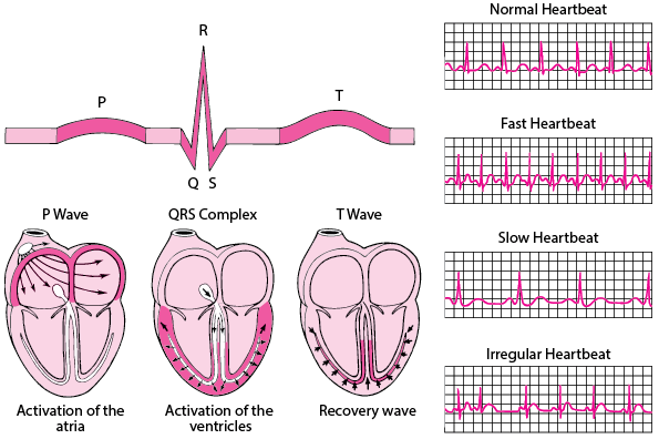
Electrocardiogram:
The electrical activity within the heart can be detected by attaching the electrodes on the arms and legs (limb leads) and at six positions on the chest (Chest leads).
Each limb and chest electrodes records slightly different electrical activity due to difference in its position, by comparing these activities with one another and with normal records, it is possible to determine
Abnormality in the conducting pathway.
Enlargement of heart.
If certain regions of the heart are damaged.
The cause of chest pain.
The instrument used is an electrocardiograph and recording of electric signals is an electrocardiogram.
The normal ECG shows three waves “P, QRS, T).

The first P wave is a small upward deflection on the ECG.
The P wave arises when the impulses from the SA node spreads over the atria.
The second wave is the QRS complex that starts with a downward deflection, continues as a large upright triangular wave and ends as a downward wave.
The QRS complex represents rapid spread of impulses from the AV node through the bundle of His and Purkinje fibres.
The third wave is dome shaped upward deflection known as the T wave. It indicates the relaxation of ventricular muscles.
Circulation of blood:
There are four types of blood circulation in the human body
Systemic circulation
Pulmonary circulation
Coronary circulation
Hepatic portal circulation
Systemic circulation:
The left ventricles pump oxygenated blood into the aorta.
From the aorta the blood divides and re-divides into arteries, arterioles and finally the blood capillaries.
The exchange of nutrients and gases occurs across the thin capillary walls.
The same set of capillaries collects deoxygenated blood from the body cells.
They unite to form venules.
Different venules come together to form veins.
The veins empty deoxygenated blood either into superior vena cava or inferior vena cava, which opens into the right atrium of the heart.
The blood circulation from the left ventricle to right atrium is called systemic circulation.
Pulmonary circulation:
This is the circulation of blood from the right ventricle of the heart to the lungs and back to the left atrium.
Right ventricles pump the blood into the pulmonary artery.
It divides into left and right pulmonary arteries and passes into the left and right lungs.
In the lungs these arteries divide and subdivide into smaller arteries, arterioles and capillaries.
The exchange of gases takes place between capillaries and the alveoli of the lungs.
Two pulmonary veins carry oxygenated blood from each lung and open into the left atrium.
Coronary circulation:
Coronary circulation is the supply of blood to the heart itself.
The right and left coronary arteries (Branches of ascending aorta) supply oxygenated blood to the heart.
The deoxygenated blood is collected by coronary veins and opens into the coronary sinus.
Hepatic portal circulation:
The venous blood from the digestive organs such as the small intestine, stomach, and pancreas is collected by a portal vein.
It empties its contents into the liver.
The portal vein is formed by different veins such as splenic vein from the spleen, inferior mesenteric from rectum and colon, superior mesenteric from small intestine, gastric vein from stomach and cystic vein from gallbladder.
In this way, blood with a high concentration of nutrients goes into the liver first.
The liver stores some of them and modifies others before they pass into general circulation.
The liver also detoxifies harmful substances, such as alcohol, that have been absorbed by the gastrointestinal tract and destroys bacteria by phagocytosis.
Hepatic artery supplies oxygenated blood to the liver, thus the oxygenated and deoxygenated blood into the liver is mixed and further collected by hepatic veins which opens into the inferior vena cava.
This circulation of blood through the liver is called hepatic portal circulation.
Commonly Asked Questions:
Write a short note on “Conduction system of Heart”.
Define “Cardiac Output”.
What two sound hearts make and why?
Write a short note on “Electrocardiogram”, give its significance.
Write a short note on,
Pulmonary Circulation.
Hepatic Circulation.
Different types of blood circulation in the human body.