Introduction:
The organ of hearing is the ear.
It is supplied by VIIIth cranial nerve (Vestibulocochlear Nerve), which is stimulated by vibrations of sound waves.
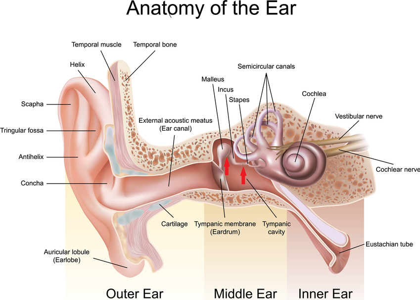
The ear is divided into three main regions.
1) External (outer ear)
2) Middle ear
3) Internal ear
1) External (Outer ear):
The external ear consists of the auricle (pinna), external auditory canal and eardrum (tympanic membrane).
The auricle is made up of elastic cartilage which is covered by skin.
The external auditory canal is a curved tube about 2.5 cm long and extends from the auricle to the eardrum.
The eardrum is a thin, semi transparent part present in between the external auditory canal and middle ear.
2) Middle ear:
The middle ear is the small cavity present in the temporal bone.
The anterior wall of the middle ear contains an opening that directly connects with the nasopharynx called the auditory tube (Eustachian tube).
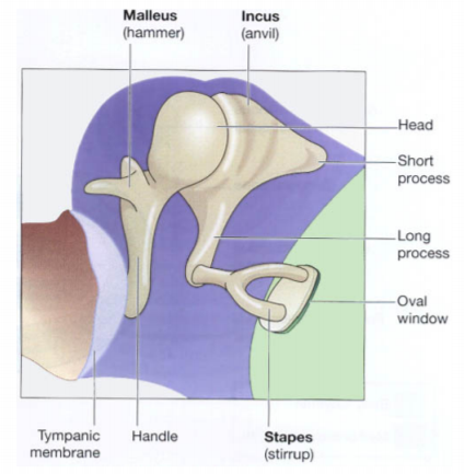
Middle ear consists of three smallest bones present in the body called auditory ossicles.
The three bones are the malleus, incus and stapes.
The one end of the malleus is attached to the internal part of the eardrum.
The other end of the malleus articulates with the body of incus.
The incus articulates with the head of the stapes.
The base of the stapes fits into the oval window.
Below the oval window is another opening called the round window.
Internal structure of Ear
3) Internal (Inner) ear
Structurally the internal ear consist of two main divisions,
a) The bony labyrinth :
It is a series of cavities present in the temporal bone.
It consists of a fluid called perilymph.
It is divided into three areas.
i) The semicircular canals -
They are anterior, posterior and lateral semicircular canals.
Each canal consists of a swollen enlargement at one end called the ampulla.
It contains receptors for equilibrium.
ii) The vestibule -
It is the oval central portion of the bony labyrinth.
It also contains receptors for equilibrium.
iii) The cochlea -
It continues with the vestibules.
It is a bony spinal canal like a snail shell and makes almost three turns around a central bony core.
The cochlea contains receptors for hearing.
b) Membranous labyrinth –
These are the series of tubes present inside the bony labyrinth.
It is lined by epithelium and contains a fluid called endolymph.
Physiology of hearing:
The auricle directs the sound waves into the external auditory canal.
These sound waves strike the eardrum and cause the eardrum to vibrate back and forth.
The vibration is transmitted from the eardrum to the malleus to the incus and then to the stapes.
The vibrations from the middle ear are transferred to the perilymph.
From the perilymph the vibrations are transmitted to the endolymph, which leads to the generation of nerve impulses.
The generated nerve impulses pass to the auditory portion of the cerebral cortex.
These impulses of hearing are interpreted by the brain.
Commonly Asked questions:
With a well labelled diagram describing the structure of the ear.
Write a short note on “Physiology of hearing”.