Joints in Human Body.
Introduction:
A joint is the place in the body where bones of the skeleton meet.
It is also called “Articulation.”
Joints allow locomotion.

Classification of Joints:
Joints can be classified as structurally or functionally.
The structural classification of joints depends on the manner in which the bones connect to each other.
The functional classification of joints depends on the nature of the movement the joints allow.
Arthrology is the branch of science that deals with the study of joints it is also called “Synosteology”.
Structural Classification of Joints:
The structural classification of joints is based on the type of tissue that binds the bones to each other at the joint.
There are three types of joints in the structural classification.
Fibrous,
Cartilaginous,
Synovial joints.
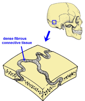
Fibrous Joints:
These are joints in which bones are joined by dense connective tissue that is rich in collagen fibers.
These joints are also called sutures.
The joints between bones of the cranium are fibrous joints.
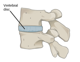
Cartilaginous Joints:
Cartilaginous joints are joints in which bones are joined by cartilage.
The joints between most of the vertebrae in the spine are cartilaginous joints.
Synovial joints:
Synovial joints are characterized by a fluid-filled space, called a synovial cavity, between the bones of the joints.
The cavity is enclosed by a membrane and filled with a fluid, called the synovial fluid, which provides extra cushioning to the ends of the bones.
Cartilage covers the articulating surfaces of the two bones, but the bones are actually held together by ligaments.
E.g. Knee joint.

Functional Classification of Joints:
The functional classification of joints is based on the type and degree of movement that they allow.
There are three types of joints in the functional classification:
Synarthrosis (Immovable).
Amphiarthrosis (Partly movable).
Diarthrosis (Freely movable).
Synarthrosis (Immovable):
Immovable joints allow little or no movement at the joint.
Most immovable joints are fibrous joints.
Besides the bones of the cranium, immovable joints include joints between the tibia and fibula in the lower leg and between the radius and ulna in the lower arm.
Amphiarthrosis (Partly movable):
Partly movable joints permit slight movement.
Most partly movable joints are cartilaginous joints.
Besides the joints between vertebrae, they include the joints between the ribs and sternum (breast bone).
Diarthrosis (Freely movable):
Movable joints allow bones to move freely.
All movable joints are synovial joints.
Besides the knee, they include the shoulder, hip, and elbow. Movable joints are the most common type of joints in the body.
Fibrous Joints:
Characteristics of the fibrous joints:
Lacks synovial cavity.
Articulating bones are joined together with help of a dense irregular connective tissue.
Offers no or very little movement.
Types of fibrous joint,
Sutures.
Syndesmoses.
Interosseous membranes.
Sutures:
It is a fibrous joint composed of a thin layer of irregular connective tissue.
It is present between the bones of the cranium.
The interlocking feature of the sutures gives them additional strength.
E.g. Coronal suture between parietal and frontal bone.
Syndesmoses:
The distance between the articulating surfaces is more.
The irregular connective tissue is denser than in sutures and is arranged in bundles.
Offers very limited movement.
e.g. The distal tibiofibular joint.
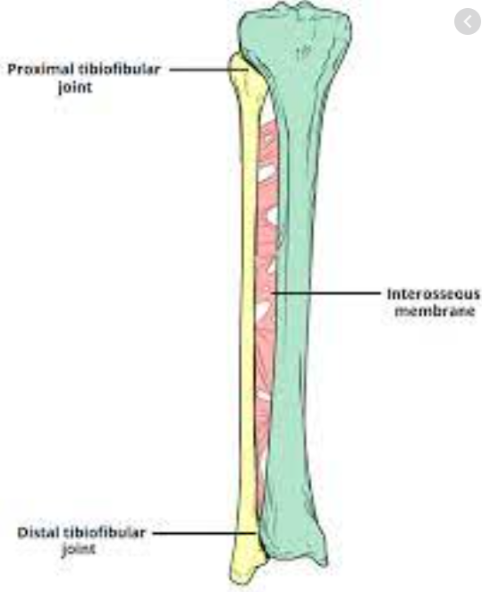
Interosseous Membrane:
It is a sheet of dense irregular connective tissues binding two neighbouring bones together.
Permits slight movement.
e.g. Between Radius and Ulna and between Tibia and Fibula.
Cartilaginous Joints:
Characteristics of cartilaginous joints:
Lacks synovial cavity.
The articulating bones are tightly connected with the help of a cartilage tissue.
Types of cartilaginous joints:
Synchondroses.
Sympahysis.
Synchondroses:
This joint has “Hyaline Cartilage” as a connecting material.
As the name indicates its a non moveable joint.
Also called “Primary Cartilaginous Joint”.
e.g. The epiphyseal plate (Growth plate) connects epiphysis and diaphysis of the growing bones.

Symphysis:
In this type of joint the ends of the articulating bones are covered by a hyaline cartilage and a broad disc of fibrocartilage connects the bones.
They alow slight movement hence amphiarthrosis in nature.
All symphysis joints occur in the midline of the body.
e.g. Pubic symphysis in hip bones.

Synovial Joints:
They show the presence of a “Synovial Cavity:.
All synovial joints are freely moveable and hence diarthrosis in nature.
A typical joint contains following structures,
Articular Cartilage.
Capsule (Ligament).
Synovial membrane.
Synovial fluid.
Intracapsular Structures.
Extracapsular Structures.
Articular Cartilage:
Made up of “Hyaline cartilage”.
It covers the ends of bones participating in the joint.
It provides a smooth and strong surface which protects the bone inside the joint.
It lacks blood supply and receives its nutrition from the synovial fluid.
Capsule:
Entire joint is covered by a sheath of fibrous tissue which holds the joint structures together.
It is loose enough to allow the movements of the joint while strong enough to avoid the injury.
Synovial Membrane:
It is made up of epithelial cells that secrete the synovial fluid.
It is present inside the capsule.
Synovial fluid:
It is a thick liquid secreted by the synovial membrane in the synovial cavity.
It provides nutrition to the structures of the joint.
Contains phagocytes that fight the infections.
Act as a lubricant.
Also act as a cushion or shock absorber.
Intracapsular Structures:
Some joints have associated structures inside the capsule e.g. Fat pads in the knee joint.
Extracapsular Structures:
The other ligaments that are associated with the capsular ligament provide stability to the joint.
Nerve and Blood Supply:
The joints receive blood supply and nerve supply to the capsule.
Types of Synovial Joints:
There are different types of synovial joints that offer different kinds of movements and articulations.
Gliding joint.
Hinge Joint.
Pivot Joint.
Condyloid Joint.
Saddle Joint.
Ball & Socket Joint.
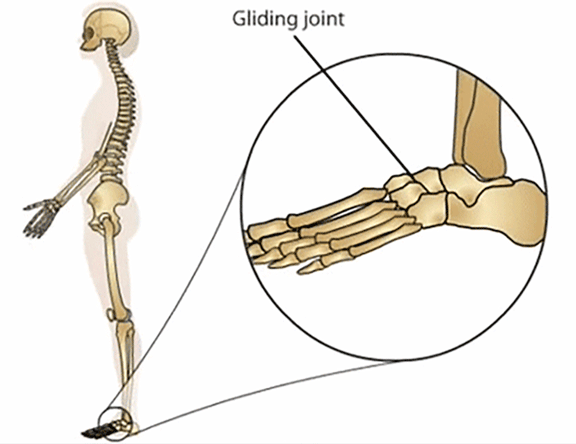
Gliding joint:
Also called “Planar Joints”.
The articulating surfaces are flat or slightly curved.
This joint allows side to side and back and forth movements between the flat surfaces of the bones.
e.g
Intercarpal joints.
Intertarsal joints.
Sternoclavicular joints.
Hinge Joint:
This joint allows an angular opening and closing motion.
One bone remains stationary while the other bone moves in an axis.
The convex surface of one bone fits in the concave surface of another bone.
They are monoaxial; they allow movement in only one direction.
e.g.
Knee joint.
Elbow joint.
Ankle joint.

Pivot Joint:
The rounded or pointed surface of one bone articulates with the ring formed by another bone.
This monoaxial allows movement in only the longitudinal axis.
e.g
Atlanto-axial joint: Between Atlas and axis which permits side to side rotation of the head.
Radioulnar joints which permit anterior and posterior movements of the palms.

Condyloid Joint:
Also called “ellipsoidal Joint”.
Here in this joint convex- oval shaped projection of one bone fits in the oval shaped depression of another bone.
Biaxial joint; movement is permitted in around two axis.
e.g.
Wrist joint.
Metacarpo-phalangeal joints.

Saddle Joint:
As the name indicates, the articulating surface of one bone is saddle shaped and the articular surface of another bone fifties in the saddle.
It is a modified condyloid joint.
Triaxial; allows movements in three axes.
e.g Carpometacarpal joint between Carpal bone: Trapezium and metacarpal of the thumb.

Ball & Socket Joint:
Here in this joint the ball-like articulating surface of one bone fits in a cup like depression of another articulating bone.
Triaxial; allows movements in three axes.
e.g.
Shoulder Joint. (The head of Humerus fits in the glenoid cavity of the scapula.)
Hip Joint (head of femur fits in the acetabulum cavity of the hip bone.)

Commonly Asked Questions:
Define and classify joints.
Describe the structure of a typical synovial joint.
Classify and describe different types of joints.
Write a note on,
Fibrous joints.
Ball and socket joints.
Pivot Joints.
Gliding Joints.
Synovial joints.